
Neurosurgical Atlas
@neurosurgatlas • 27,001 subscribers
Dr. Aaron Cohen-Gadol’s most respected & popular collection of advanced microneurosurgical techniques in the world. https://t.co/VyskBO5DCg…
Videos

In this operative video, Dr. Cohen demonstrates the telovelar approach for resection of a large hypervascular vermian hemangioblastoma. The interface between the hemangioblastoma and surrounding tissue is developed with coagulation and sharp dissection. As the dissection advances deeper, bleeding risk increases, so any capsular entry is controlled with further coagulation. The tumor is operated on from deep to superficial to protect the floor of the fourth ventricle while identifying larger anterior vessels. Only vessels clearly entering the tumor are coagulated, preserving cerebellar vessels and maintaining hemostasis near the fourth ventricular floor. Learn more here: #Neurosurgery #Hemangioblastoma #DrCohen #SurgicalExcellence
Neurosurgical Atlas144,636 views • 1 month ago

0:59
Sensitive content
This media may contain sensitive content.
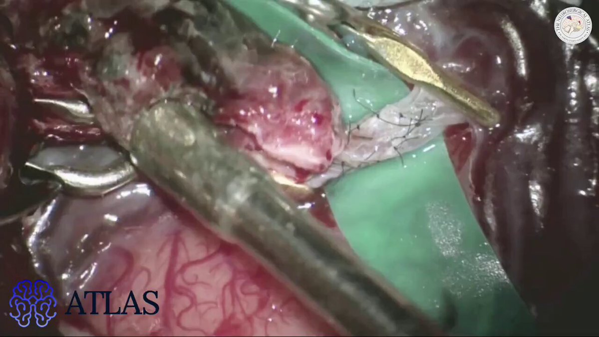
In this operative video, Dr. Cohen demonstrates a posterior callosotomy/trans-splenium approach for resection of a giant posterior biventricular epidermoid tumor. The endoscope is introduced to inspect posterior areas that could not be seen through the transcallosal approach. Under endoscopic visualization, residual tumor fragments are removed while bimanual dissection is maintained. The capsule is removed as safely as possible to reduce the chance of future recurrence, though its dense adherence to the surrounding ventricles makes gross total resection unsafe. Learn more at: #Neurosurgery #EpidermoidTumor #DrCohen #SurgicalExcellence
Neurosurgical Atlas199,130 views • 2 months ago
1:00
Sensitive content
This media may contain sensitive content.
In this operative video, Dr. Cohen demonstrates the frontotemporal approach for STA-MCA bypass and proximal M2 occlusion of a giant MCA aneurysm. After the bypass anastomosis is completed, the temporary clips are removed and mild bleeding from the site confirms graft patency. Cotton is used to reinforce the anastomotic site, and Doppler ultrasonography verifies flow through the donor vessel. A temporary clip is then placed proximal to the aneurysm, with intraoperative angiography confirming minimal aneurysm filling and preserved bypass flow before permanent clip placement is performed. Learn more here: #Neurosurgery #MCAAneruysm #DrCohen #SurgicalExcellence
Neurosurgical Atlas87,800 views • 3 months ago
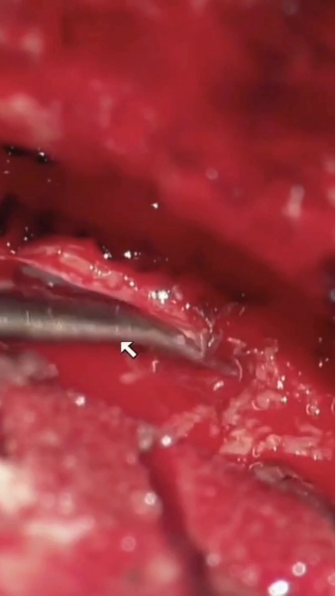
In this operative video, Dr. Cohen demonstrates resection of a cavernous sinus meningioma through a frontotemporal approach. The operation proceeds through the middle fossa, where the fibrous tumor is entered inferiorly and internally debulked to create working space. As the mass is reduced, the dissection follows the lateral wall of the cavernous sinus, with continued attention to the trigeminal nerve branches as they are identified and preserved. Further opening of the lateral cavernous sinus wall allows additional tumor removal until cavernous sinus venous bleeding defines the limit of safe resection. Start your trial with the Neurosurgical Atlas today to gain access to our library of operative videos.
Neurosurgical Atlas13,173 views • 14 days ago

In this operative video, Dr. Cohen demonstrates staged retromastoid and pterional approaches for resection of a petroclival epidermoid tumor. Careful attention is paid to the fourth cranial nerve as it enters the tentorium near the petrous apex. Tumor along the posterior basal temporal lobe is removed through the supratrochlear working corridor. The portion anterior to the midbrain is dissected while the contralateral oculomotor nerve is identified and protected, permitting maximal safe removal of tumor extending toward the medial temporal lobe. Learn more here: #Neurosurgery #EpidermoidTumor #DrCohen #SurgicalExcellence
Neurosurgical Atlas13,211 views • 19 days ago

In this operative video, Dr. Cohen demonstrates the supracerebellar transtentorial approach for resection of a medial tentorial meningioma. After devascularization, the tumor becomes more gelatinous and is removed with suction while posterior cerebral artery branches are protected to prevent occipital ischemia. The tumor-involved tentorium is then pulled into the resection cavity after its medial attachment is released. Smaller arachnoid bands are divided, the field is cleared, and the affected tentorial segment is removed. Final inspection shows no obvious residual tumor in the field. Learn more here: #Neurosurgery #Meningioma #DrCohen #SurgicalExcellence
Neurosurgical Atlas19,703 views • 1 month ago

In this operative video, Dr. Cohen demonstrates the frontotemporal approach for clip ligation of a large calcified MCA aneurysm. A tandem clipping strategy is used with a straight fenestrated clip followed by a straight clip to close the distal and proximal aneurysm neck. Although ICG initially suggests no filling, fluorescein angiography demonstrates persistent aneurysm filling. Additional straight and fenestrated clips are placed, but calcification prevents complete closure and the sac continues to fill. Ultimately, multiple clips are required to fully exclude the partially calcified atherosclerotic aneurysm. Learn more here: #Neurosurgery #Aneurysm #DrCohen #SurgicalExcellence
Neurosurgical Atlas13,427 views • 1 month ago

In this operative video, Dr. Cohen demonstrates a suboccipital craniotomy for proximal-to-distal PICA end-to-side anastomosis for treatment of a calcified fusiform distal PICA aneurysm. The distal temporary clip is removed, revealing backflow through the aneurysm, so a permanent curved clip is placed on the distal aneurysm segment to prevent continued filling. The remaining temporary clips are removed sequentially, with minimal bleeding at the anastomosis. Micro-Doppler and fluorescein angiography confirm good flow through the vessels, no aneurysm filling, and preserved perfusion of the fourth ventricular floor. Learn more at: #Neurosurgery #Aneurysm #DrCohen #SurgicalExcellence
Neurosurgical Atlas19,378 views • 2 months ago

In this operative video, Aaron Cohen-Gadol, MD demonstrates the contralateral cross-court approach for clip ligation of a right P1 aneurysm through a left-sided pterional craniotomy. The basilar artery, its bifurcation, and the contralateral P1 segment are exposed, revealing a perforating vessel arising from the neck of the P1 aneurysm. The cross-court approach is effective in this case, as the anatomy provides favorable visualization for circumferential dissection of the neck. A temporary clip is placed on the basilar trunk to allow controlled manipulation of the neck for definitive clip application. The narrow working space medial to the carotid artery is used to inspect the perforating vessels. A long straight clip is then applied to collapse the aneurysm neck, providing adequate visualization around the blades to preserve the perforators. Learn more here: #Neurosurgery #AneuerysmClipping #DrCohen #SurgicalExcellence
Neurosurgical Atlas26,296 views • 9 months ago
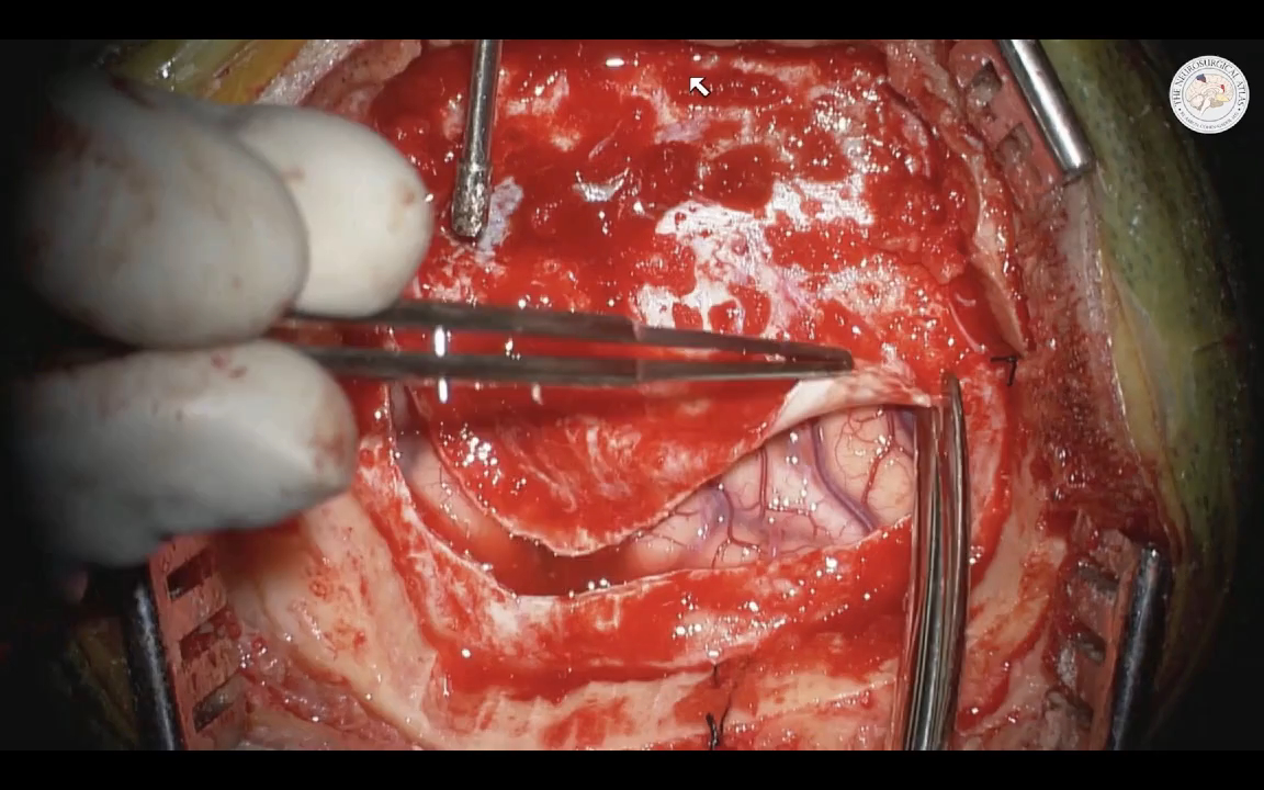

Wishing you all a joyful Thanksgiving! With 70,000+ global members, the Neurosurgical Atlas impacts daily patient care. Exciting things await in the coming year, including the Atlas Intelligence platform. Inspired by requests from many of you, enjoy the art of carving a turkey.
Neurosurgical Atlas24,341 views • 2 years ago

1:00
Sensitive content
This media may contain sensitive content.
No more content to load