
sergio serrano belmar
@sserranobmsk • 2,796 subscribers
MSK Radiologist Real Madrid - MDAnderson - Ruber Internacional - Federación Española de Rugby
Shorts

Videos

Diferential diagnosis of insertional plantar fasciitis: part one A patient with plantar pain that had been going on for months was diagnosed with fasciitis, with no response to physiotherapy treatment, two corticosteroid infiltrations and three PRP infiltrations. She reports intermittent plantar and medial plantar pain, sometimes being more intense early in the day. Minimal non-painful thickening at the origin of the plantar fascia is identified sonographically. On clinical examination and ultrasound, there was pain along the posterior tibial tendon, with a positive double heel-rise test, so ultrasound-guided infiltration was performed with 1 cc of triamcinolone and 1 cc of 2% lidocaine in its sheath, with partial resolution of the symptomatology. Lumbar exploration was performed, with pain due to compression of the S1 sacral foramina that correlated with the dermatome of the sensitive area at the level of the heel (dependent on the branch of the tibial nerve), so transforaminal infiltration was performed with 1 cc of triamcinolone and 1 cc of 2% lidocaine, with complete resolution of symptoms.
sergio serrano belmar36,354 次观看 • 2 年前

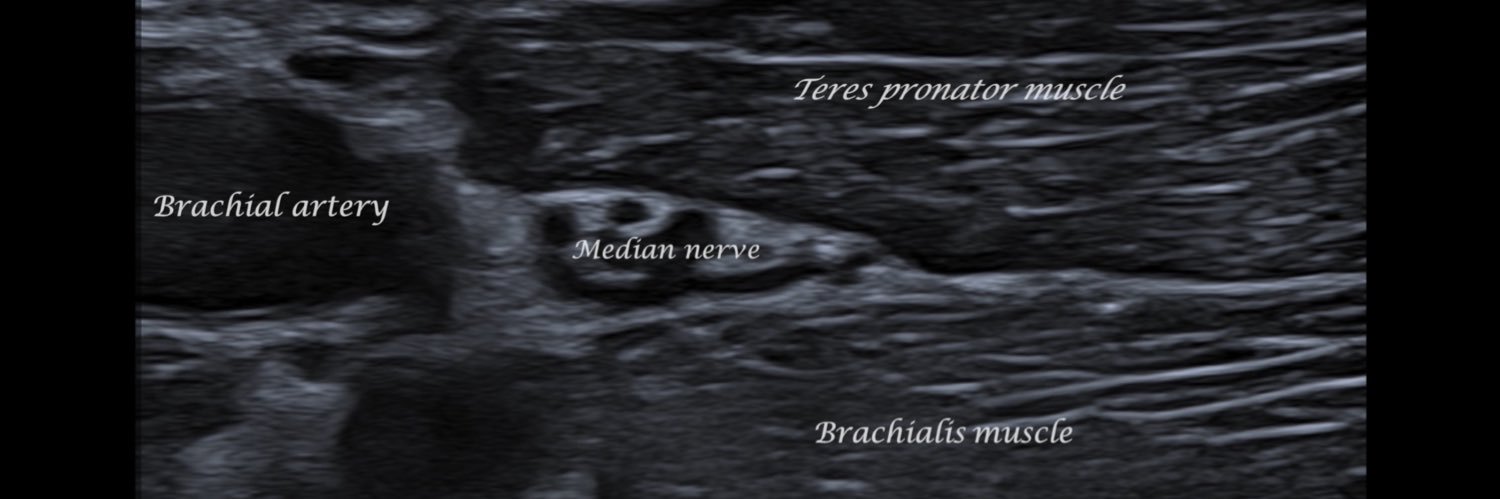
Abdominal wall pain: differential diagnosis A 28-year-old soccer player presented with right periumbilical pain of 2 months' duration. An MRI was performed, which showed no abnormalities in the abdominal wall. He was diagnosed with anterior cutaneous nerve entrapment syndrome (ACNES), and perineural injection was performed, with no improvement in symptoms. He came to the clinic for a second opinion, presenting with tenderness on palpation of the middle third of the rectus abdominis muscle, which increased with trunk resistance flexion. The pain decreased after this test was performed after muscle inhibition. An ultrasound assessment was completed, revealing no abnormalities in the discomfort, but evidence of increased thickness and decreased echogenicity of the tendon at its origin in the pubic ramus, associated with significant pain on sonopalpation, related to tendinosis. An evaluation was completed with a thoracic spine examination, revealing significant pain at the level of the spinous process of T8 (the rectus abdominis muscle is innervated by the T7-T12 thoracoabdominal nerves). It was decided to treat the patient with ultrasound-guided injections around the rectus abdominis tendon with 1 cc of triamcinolone and 1 cc of 2% lidocaine, and periradicular injections of the right T8 muscle with 2 cc of betamethasone and 1 cc of 2% lidocaine. The symptoms resolved immediately, and the patient did not reappear during the following two months of follow-up.
sergio serrano belmar18,963 次观看 • 1 年前

The usefulness of the fibrillar pattern and pennation angle in the ultrasound diagnosis of muscle injury A 17-year-old patient who, while braking while playing football, feels a sharp pain in the anterolateral region of the proximal third of the thigh that prevents him from continuing to play sports. Typically, the axial plane (the short axis of the muscle) is used to study fibrillar tears, with a high diagnostic sensitivity. However, on many occasions, it can be easier to scan through the long axis of the muscle to visualize the different muscle fascicles and the pennation angle in their entirety, being able to make the diagnosis by observing the interruption and loss of tension of the muscle fascicles associated with the loss of the normal pennation angle. In the present case, the normal fibrillar pattern is initially visualized in the medial region of the rectus femoris muscle until the presence of a fibrillar tear in the posterolateral myofascial junction of its proximal third, where there is a sudden interruption of the fibrillar pattern due to the presence of an intramuscular hematoma.
sergio serrano belmar13,140 次观看 • 1 年前

Trigger finger: ultrasound diagnosis and treatment Patient with stiffness in the first finger of the right hand, especially in the morning, which is accompanied by a sensation of clicking or crunching when moving the finger, associated with sensitivity and a lump in the palm at the level of the metacarpophalangeal joint. An ultrasound study shows a diffuse increase in the thickness of the flexor tendon, observing the tendon snapping at the level of pulley A1 in a dynamic study when the tendon slides. Initially, infiltration of 1 cc of trigon and 1 cc of 2% mepivacaine into the tendon sheath is performed under ultrasound control to subsequently proceed to fenestration of the pulley until the tendon is released. Sometimes, it can be associated with physiotherapy to unload the muscles in the forearm, performing proximal peritendinous infiltration of the first finger's flexor muscle to prevent the pathology's recurrence.
sergio serrano belmar11,965 次观看 • 1 年前

Adhesive capsulitis of the hip: diagnosis A 55-year-old patient with gluteal pain of 6 months' duration associated with decreased range of motion of the hip joint. An AMRI is requested to rule out a tear of the acetabular labrum. The study shows an increase in thickness of the lower and upper bands of the iliofemoral ligament (measuring 13 and 15 mm, respectively) associated with pericapsular edema, consistent with adhesive capsulitis. An ultrasound-guided intra-articular infiltration of 1 cc of Trigon and 1 cc of 2% mepivacaine is performed, and the pain is entirely resolved after five days. Treatment is associated with physiotherapy and rehabilitation, and the range of joint mobility recovers after three weeks. In the article “Capsular ligaments of the hip: anatomic, histologic, and positional study in cadaveric specimens with MR arthrography” (doi: 10.1148/radiol.12111320), the average thickness of the capsular ligaments of the hip joint is described.
sergio serrano belmar11,323 次观看 • 1 年前

dry needling: MRI findings Patient with pain in the long head of the biceps femoris muscle and a suspected fibrillar tear, for whom an MRI of the thigh was requested. The MRI shows a patchy increase in signal in the distal third of the long head of the biceps associated with hyperintense linear paths, which correlate with the history of dry needling performed three days before the radiological study. Dry needling is an invasive technique used in physiotherapy to eliminate muscle pain points (or trigger points), which can be generated due to overload. It is imperative not to do this treatment during the five days before performing an MRI because it can make the diagnosis of muscle injury difficult or impossible, mainly grade I. How many routes do you see in the video?
sergio serrano belmar12,609 次观看 • 2 年前
没有更多内容可加载