正在加载视频...

视频加载失败

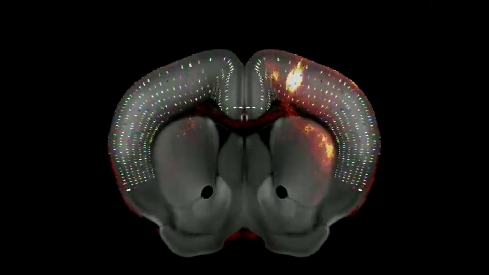
Freely access electrophysiological, morphological, and transcriptomic data measured from thousands of individual mouse & human brain cells via the Allen Cell Types Database: #openscience #SfN23

30,850 次观看 • 2 年前 •via X (Twitter)

0 条评论

暂无评论

原始帖子的评论将显示在这里
相关视频

New Cell paper from Bergles lab at Johns Hopkins just built the most comprehensive map of brain myelin ever made — every oligodendrocyte, across the entire mouse brain, across the lifespan. The scale: >10 million cells per brain, terabyte-scale 3D lightsheet volumes, registered to the Allen Brain Atlas across 417 regions from 2 months to 2+ years of age. The technical stack: Custom tissue clearing (CUBIC-L + SHIELD + uRIMS with 40% urea) to preserve endogenous fluorescence. 3D Mask R-CNN for instance segmentation — not just semantic, instance — so it can distinguish individual cells within dense clusters at scale via overlapping sliding windows. Vision Transformer to classify newly-formed vs. mature oligodendrocytes using soma morphology. All cross-referenced against Allen ISH transcriptomics and MICrONS serial EM. What they found: Oligodendrocyte density varies 10,000-fold across brain regions. Left-right hemispheres: r=0.99. Sex: no significant difference. Strain: matters. The brain never stops myelinating. New oligodendrocytes are still being generated in 2-year-old mice. Prefrontal cortex L6 shows the fastest rates of new myelination into old age — the circuits for executive function keep rewiring throughout life. After demyelination, L4 sensory cortex is the most resilient — oligodendrocytes survive at higher rates. The hippocampus loses nearly everything and barely recovers. Degree of injury doesn't predict rate of recovery. These are independent axes. The Alzheimer's result is the most surprising: Dense-core plaques dominate in cortex and hippocampus. Diffuse/small-core plaques dominate in white matter fiber tracts. Old assumption: diffuse plaques are "less toxic." The data says the opposite — small plaques in fiber tracts cause more myelin loss per plaque than dense-core plaques in gray matter. Plaque load and oligodendrocyte loss are essentially uncorrelated (ρ=0.22). The damage is plaque-type and location specific, not load-dependent. For MS and AD research: you can't read off white matter injury from gray matter plaque burden. The pathology in fiber tracts is running on different rules. Data: Paper:

Bo Wang

24,777 次观看 • 6 个月前

The most detailed 3D reconstruction of a cell ever created. Blows my mind every time. But what exactly are we looking at here? The average human cell contains: ~ 15-20 total distinct organelle types, totalling between ~1-10 million working together per cell. All these nano-machines in the cell are made up of proteins. ~ 8,000-10,000 distinct types of unique proteins, adding up to between 40 million - 10 trillion total proteins making up all those cellular systems. ~ 10,000 - 15,000 distinct types of RNA shuttling information around the cell, totalling up to ~10 million RNA molecules moving around the cell simultaneously. ~ Billions of Lipid molecules packed together into the cell membrane, which is also packed tightly with millions more protein-based nano-machines. And let's not forget billions of lines of DNA information to build and run it all. That's TRILLIONS of of individual molecular pieces working together to make a single cell function. That means there is more complexity in a single cell than humanity's largest cities. And people still believe this wasn't Divinely Designed. This is God's Glory on Display. But to make the point. A cell couldn't have evolved from some nebulous simpler "protocell" because even the simplest cells still require massive complexity. The "simplest" cell ever created was engineered by scientists knocking out pieces of a functional cell until it stopped functioning. Here is what they found is the absolute necessary minimal requirements of a cell to function: - Over ~531,000 lines of coded DNA information - 473 total genes to create hundreds of unique protein products (they later added 19 genes back in because the cell was so weak) - Hundreds of thousands of total proteins all working together - Extensive regulatory networks guiding all these interactions If the cell doesn't have all these systems in place, from the start... it doesn't live. Cell rely on an intricate network of complex systems, which are themselves built from complex interconnected pieces woven together into an incomprehensibly complex web of functionilty. Only intelligence has ever been observed creation vast interconnected systems like this. Life was clearly Created. It couldn't happen any other way.

Divinely Designed

166,503 次观看 • 3 个月前

🚨 Scientists discover wisdom teeth contain stem cells capable of repairing the heart, brain, and bones. Wisdom teeth contain dental pulp, a soft connective tissue threaded with blood vessels and nerves. Inside that pulp lives a dense population of mesenchymal stem cells, a class of undifferentiated cells that researchers classify as among the most therapeutically valuable biological material a human body produces. These are not ordinary cells maintaining routine tissue. They are blueprint cells, capable of receiving chemical signals from damaged environments and reshaping themselves into whatever the body needs most, neurons, cardiomyocytes, osteoblasts, even hepatic cells under the right conditions. The brain operates under a brutal rule: most of its neurons do not regenerate after damage. A stroke, a traumatic injury, a neurodegenerative disease removes cells the brain cannot replace through normal biological processes. Researchers have spent decades attempting to solve this through synthetic means, engineered cell therapies, growth factor injections, gene editing approaches that cost extraordinary resources and produce inconsistent results. What dental pulp stem cells demonstrated in laboratory conditions is that they can migrate toward neural damage sites, integrate with existing tissue architecture, and begin producing neurons and glial support cells. The mechanism involves neurotrophic factor secretion, essentially the cells releasing signaling proteins that stimulate the surrounding neural environment to repair itself from within. Cardiac muscle operates under a similarly unforgiving rule. After a heart attack, the dead muscle tissue becomes fibrotic scar material. The heart compensates by making surviving muscle work harder, a process that gradually leads to enlargement, weakening, and eventual failure. Dental pulp stem cells introduced into cardiac tissue in multiple studies produced measurable reductions in scar formation and demonstrated the ability to differentiate into functional cardiomyocytes, beating in synchrony with native heart cells. Some studies recorded improved ejection fraction in animal models, the core measurement of how effectively the heart pumps blood. Bone regeneration represents the most clinically advanced application already moving toward human trials. Dental pulp stem cells express high levels of osteogenic markers and respond rapidly to bone morphogenetic proteins, the chemical messengers that trigger skeletal repair. Their application in craniofacial reconstruction, spinal fusion, and long bone defect repair is being studied across multiple institutions simultaneously. What separates these cells from other stem cell sources is the combination of accessibility and biological youth. Bone marrow aspiration requires sedation and produces significant post procedure pain. Umbilical cord blood requires planning around birth. Wisdom teeth emerge between 17 and 25, during peak cellular vitality, and come out during a procedure most people already schedule. The extraction window is permanent. Once the teeth are gone and the pulp degrades, that specific population of young, highly potent cells is irretrievable from that individual. Cryogenic preservation protocols now exist that maintain dental pulp stem cell viability for over two decades. Several countries have commercial dental stem cell banks operating with the same institutional model as cord blood banking, long term frozen storage, indexed against future therapeutic need. The science supporting the value of preservation is no longer speculative. What lags behind is public awareness and clinical infrastructure in markets where this remains obscure. The wider pattern is worth recognizing. Medicine has repeatedly discovered that profound biological tools were present in tissues it previously categorized as vestigial, unnecessary, or inconvenient. The appendix was considered evolutionary junk for over a century before researchers identified its role in gut microbiome preservation. Wisdom teeth carried the same dismissal, a developmental relic from ancestors who needed extra molars for coarse diets, relevant only in their capacity to cause orthodontic problems. The pulp inside them was never junk. It was a repair system the body built during youth and stored in one of the most protected anatomical locations, surrounded by enamel, the hardest substance the human body produces. Evolution rarely wastes that kind of architecture.

The Curious Tales

24,267 次观看 • 5 个月前

To replace animal testing with AI, we need MASSIVE human datasets. Today, we're thrilled to share Axiom's new data exploration tool, providing the ability to visually explore the world's largest primary human liver toxicity dataset. Built with Axiom's proprietary wetlab protocols, our dataset includes detailed liver toxicity profiles for over 100,000 distinct molecules. The key to this dataset is our ability to do high-throughput, multiplexed high-content screening with primary human liver cells. Traditionally, toxicity assays either sacrifice throughput or sacrifice biological relevance (using easy-to-grow immortalized cell lines instead of real human cells). We managed to combine throughput, physiological relevance, and multiplexing in one platform. The assays run in a high throughput format using automation, meaning thousands of compound-dose conditions can be tested in one experiment. We achieved this using pooled primary human hepatocytes, which are often fragile and expensive. By systemizing our automation and quality control processes, we were able to run over 120+ batches on the same donor pool with incredible reproducibility and consistency. We did this while integrating many readouts per well, whereas many existing toxicity assays only do a single readout. Our multiplexed approach provides far more data per experiment enabling us to measure 10-20 different toxicity phenotypes such as apoptosis, necrosis, mitochondrial fission, endoplasmic reticulum stress, stress granule formation, microtubules, and more all from a single well on a 384-well plate! The combination of scale, high content information, and data quality is exactly what is needed to train highly accurate AI models in biology. If you're interested, please explore the dataset in the comments below and let me know if you want to chat about the details!

Brandon White

25,117 次观看 • 1 年前