Peter M. Lawrence's banner
Peter M. Lawrence's profile picture

Peter M. Lawrence

@PeterMLawrence142,387 subscribers

🧠✍🏻 Certified medical illustrator @barrowneuro

Shorts

I’m very excited about this anatomy overlay test print. The final product will have layers of anatomy, from skin down to a midsagittal section through the skull, brain and spine, with labels

I’m very excited about this anatomy overlay test print. The final product will have layers of anatomy, from skin down to a midsagittal section through the skull, brain and spine, with labels

511,438 views

The posterior inferior cerebellar artery (PICA) has five segments: P1, anterior medullary: located in front of the medulla (blue) P2, lateral medullary: runs alongside the medulla and ends at the origin of the glossopharyngeal, vagal, and accessory nerves (purple) P3, tonsillomedullary: runs around the caudal half of the cerebellar tonsil (green) P4, telovelotonsillar: runs in the cleft between the tela choroidea and the inferior medullary velum, and the superior pole of the cerebellar tonsil (orange) P5, cortical: distributed to the cerebellar surface (yellow) Illustrated for Michael T. Lawton, MD ‘s 7 Cavernomas #Neuroscience #MedTwitter #NeuroTwitter Oren Gottfried, MD The Innovation | Medicine

The posterior inferior cerebellar artery (PICA) has five segments: P1, anterior medullary: located in front of the medulla (blue) P2, lateral medullary: runs alongside the medulla and ends at the origin of the glossopharyngeal, vagal, and accessory nerves (purple) P3, tonsillomedullary: runs around the caudal half of the cerebellar tonsil (green) P4, telovelotonsillar: runs in the cleft between the tela choroidea and the inferior medullary velum, and the superior pole of the cerebellar tonsil (orange) P5, cortical: distributed to the cerebellar surface (yellow) Illustrated for Michael T. Lawton, MD ‘s 7 Cavernomas #Neuroscience #MedTwitter #NeuroTwitter Oren Gottfried, MD The Innovation | Medicine

42,957 views

#Throwback: still one of my favorite books—Wendell J. S. Krieg’s “Human Brain in Diachrome” has unique, stunning illustrations printed on clear slide overlays that depict neuroanatomy, tracts, and nuclei. I haven’t been able to find more copies online. If anyone knows where you can get them, please let DM me. Dr. Wendell Krieg (1906–1997) was an American neuroanatomist, educator, and scientific illustrator whose work helped bridge the traditions of classical neuroanatomy and modern neuroscience. Trained at the University of Wisconsin and later a professor of anatomy at Northwestern University Medical School, Krieg devoted much of his career to studying and teaching the structural organization of the brain. A skilled draftsman, he produced clear schematic illustrations of neural pathways and cortical connections that became widely used in medical education. In 1947 he founded the Cajal Club, an international society dedicated to honoring the legacy of Santiago Ramón y Cajal and advancing the study of the nervous system. Through his research, teaching, and visualizations of brain circuitry, Krieg helped preserve the intellectual lineage of Cajal’s neuron doctrine while shaping mid-twentieth-century approaches to functional neuroanatomy. #medicalillustration

#Throwback: still one of my favorite books—Wendell J. S. Krieg’s “Human Brain in Diachrome” has unique, stunning illustrations printed on clear slide overlays that depict neuroanatomy, tracts, and nuclei. I haven’t been able to find more copies online. If anyone knows where you can get them, please let DM me. Dr. Wendell Krieg (1906–1997) was an American neuroanatomist, educator, and scientific illustrator whose work helped bridge the traditions of classical neuroanatomy and modern neuroscience. Trained at the University of Wisconsin and later a professor of anatomy at Northwestern University Medical School, Krieg devoted much of his career to studying and teaching the structural organization of the brain. A skilled draftsman, he produced clear schematic illustrations of neural pathways and cortical connections that became widely used in medical education. In 1947 he founded the Cajal Club, an international society dedicated to honoring the legacy of Santiago Ramón y Cajal and advancing the study of the nervous system. Through his research, teaching, and visualizations of brain circuitry, Krieg helped preserve the intellectual lineage of Cajal’s neuron doctrine while shaping mid-twentieth-century approaches to functional neuroanatomy. #medicalillustration

11,013 views

I had the honor of observing hundreds of original watercolor and airbrush pieces by father & son medical illustrators, Gerhart & Stephan Spitzer. Stephan generously invited me to stay with him and his wife in Frankfurt Germany for a few days. He and his father have produced numerous anatomical and surgical atlases published by Thieme Medical Publishers. His archive contains thousands of beautifully rendered artworks rendered in pen & ink, graphite, watercolor, and many other media. It was such a great opportunity—thank you, Stephan. #medicalillustrator #anatomy

I had the honor of observing hundreds of original watercolor and airbrush pieces by father & son medical illustrators, Gerhart & Stephan Spitzer. Stephan generously invited me to stay with him and his wife in Frankfurt Germany for a few days. He and his father have produced numerous anatomical and surgical atlases published by Thieme Medical Publishers. His archive contains thousands of beautifully rendered artworks rendered in pen & ink, graphite, watercolor, and many other media. It was such a great opportunity—thank you, Stephan. #medicalillustrator #anatomy

21,862 views

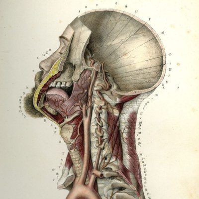
Head & neck anatomy 🧠 #neurotwitter

Head & neck anatomy 🧠 #neurotwitter

11,173 views

The posterior cerebral artery (PCA) has five segments: P1, pre-communicating segment, originates at the end of the basilar artery & terminates at the posterior communicating artery (green) P2, post-communicating segment, begins after the posterior communicating artery (PCOM) and loops around the midbrain & terminates at the origin of the lateral occipital artery as it enters the quadrigeminal cistern (blue) P3, quadrigeminal segment, which runs through the quadrigeminal cistern in a posteromedial direction (purple) P4, cortical segment, located within the sulci of the occipital lobe (yellow) P5, terminal branches (orange) #neuroeducacion #MedTwitter Oren Gottfried, MD The Innovation | Medicine

The posterior cerebral artery (PCA) has five segments: P1, pre-communicating segment, originates at the end of the basilar artery & terminates at the posterior communicating artery (green) P2, post-communicating segment, begins after the posterior communicating artery (PCOM) and loops around the midbrain & terminates at the origin of the lateral occipital artery as it enters the quadrigeminal cistern (blue) P3, quadrigeminal segment, which runs through the quadrigeminal cistern in a posteromedial direction (purple) P4, cortical segment, located within the sulci of the occipital lobe (yellow) P5, terminal branches (orange) #neuroeducacion #MedTwitter Oren Gottfried, MD The Innovation | Medicine

16,172 views

Videos

No more content to load