Loading video...

Video Failed to Load

Go Home

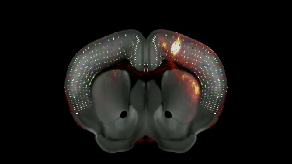
New microscopy technology enables nanoscale imaging of centimetre-scale tissues, making it possible to image the entire expanded mouse brain at cellular and subcellular resolution. #ReviewedPreprint

35,249 views • 2 years ago •via X (Twitter)

0 Comments

No comments available

Comments from the original post will appear here

Related Videos

I've been editing this article about "brain mapping" and connectomics, and I'm just stunned by how quickly the cost estimates to map, say, a mouse brain have plummeted in just the last couple years. It actually seems feasible that we could map the entire human brain -- all 86 billion neurons, and their connections -- in this lifetime. In the 1970s, Sydney Brenner started mapping all the connections between neurons in C. elegans. His team sliced the worm into thin pieces, took photos using an electron microscope, and manually traced and reconstructed each synapse for 302 neurons total. This project took more than a decade of work, and it cost about $16,500 to reconstruct each neuron. Scaling this up to a human brain boggles the mind. Electron microscopy remained the norm in connectomics for decades, because it was the only option available to see synapses at a resolution high enough to be able to trace their paths. Each electron microscope costs several hundreds of thousands of dollars, though, and you need lots of them to map even a mouse brain in a reasonable timeframe. In 2023, the Wellcome Trust released a report estimating how long, and how expensive, it would be to map the mouse connectome (~70M neurons). They estimated that imaging alone would cost $200-300M, and that proofreading (or ensuring that traces between neurons are correct) would cost $7-21 BILLION. (A human can only manually trace about 1 mm of neuron per hour.) Also, the images would occupy about 500 petabytes of data, and getting those data would require 20 electron microscopes running in parallel for about 5 years, continuously. They estimated the whole project would take about 17 years of work. This is, understandably, insane. But now it seems like there's an actual path toward mapping the full mouse brain in about five years for ~$100M dollars. There have been three major breakthroughs in the last year or so: 1/ Expansion microscopy, first developed in 2015, showed that it's possible to "enlarge" the brain by about 5x using a swellable polymer. But an improved method increases this number to >20x expansion, meaning we can now expand brains and image neurons much more easily using cheap light microscopes, rather than expensive electron ones. 2/ E11 Bio (a nonprofit research org) developed protein barcodes that get delivered into brain tissue; each neuron gets a unique combination of barcodes. These cells are then stained with colorful antibodies, which stick to a matching protein barcode, causing each neuron to light up in a distinct color. This makes tracing neurons so much easier. 3/ Google Research released PATHFINDER this May, an AI-based neuron tracing tool that can proofread about 67,200 cubic microns of brain tissue per hour, with very high accuracy. It works on electron micrographs, but something similar could be presumably be developed for the E11 / colorful tag approach. This is an extremely exciting time for neuroscience. (C. elegans connectome below.)

Niko McCarty.

66,819 views • 7 months ago

🔬 Why Perfect Visualization of Stents and Calcified Plaques Requires the Highest Spatial, Temporal and Contrast Resolution: The Case for Ultra-High-Resolution with Photon Counting CT Image: a close look at a RCA stent with very mild intimal hyperplasia. Seeing is everything in coronary imaging. Yet when it comes to stents and heavily calcified plaques, even the best conventional CT systems hit their physical limits — blooming, partial volume effects, and metal artifacts blur what truly matters. That’s where Ultra-High-Resolution Photon Counting CT (PCCT) changes the game. 🧠 Why it matters: Conventional CT often overestimates stenosis in calcified vessels and hides in-stent lumen due to blooming and noise. This leads to diagnostic uncertainty, unnecessary invasive testing, and poor evaluation of stent patency or neo-atherosclerosis. ⚡ What PCCT adds: Ultra-high spatial resolution (0.1 mm voxels) sharply delineates stent struts, plaque borders, and residual lumen. True spectral resolution minimizes blooming and metal artifacts, enhancing visualization even in highly calcified segments. Improved contrast-to-noise enables clear lumen assessment at lower doses. Quantitative, energy-resolved data provide material differentiation between calcium, metal, and contrast — something previously unthinkable in CT. In short, accurate visualization of stents and calcified plaques demands Photon Counting CT — not just for sharper images, but for better diagnostic confidence and patient management. The era of artifact-free coronary imaging has begun — and it’s photon-counting powered. ⚡❤️ #PCCT #CardiacImaging #CoronaryCT #StentImaging #CalciumScore #PhotonCountingCT #PrecisionImaging #CardiovascularImaging #RadiologyInnovation #yesCCT

Dr. Filippo Cademartiri

11,093 views • 6 months ago