正在加载视频...

视频加载失败

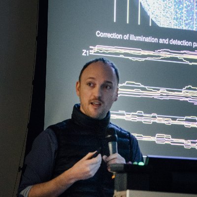
We present #Zebrahub: a timecourse atlas of zebrafish embryonic development, combining #scRNAseq time-course data with #lightsheet live imaging. Explore our seq. and imaging datasets interactively at 1/n
86,052 次观看 • 3 年前 •via X (Twitter)

25 条评论

Loïc A. Royer 💻🔬⚗️ 的头像
Loïc A. Royer 💻🔬⚗️3 年前

All datasets, instrument designs, software packages, protocols are accessible for download at to facilitate the reproducibility and accessibility of our research. We are uploading some datasets & cleaning repos, please be patient with us ;-) 2/n

Loïc A. Royer 💻🔬⚗️ 的头像
Loïc A. Royer 💻🔬⚗️3 年前

The scRNAseq dataset consists of a single-embryo high-quality #scRNAseq timecourse dataset for zebrafish post-gastrulation development (10, 12, 14, 16, 19, 24 hpf). In addition, we also provide 2, 3, 5, and 10 days post-fertilization data. 3/n

Loïc A. Royer 💻🔬⚗️ 的头像
Loïc A. Royer 💻🔬⚗️3 年前

Our optimized single-embryo cell dissociation protocol lets us do a detailed analysis of gene expression variance across sibling embryos. Interestingly, the gene expression distributions across siblings reach a point of minimal gene expression variability at 19 hpf. 4/n

Loïc A. Royer 💻🔬⚗️ 的头像
Loïc A. Royer 💻🔬⚗️3 年前

The scRNAseq data captures the main temporal and anatomical developmental features of vertebrate embryogenesis. From the establishment of the nervous system to mesenchyme lineages. We identified a total of 200 cell types across all 10 developmental stages. 5/n

Loïc A. Royer 💻🔬⚗️ 的头像
Loïc A. Royer 💻🔬⚗️3 年前

We also applied the RNA velocity method to our data and obtain a striking 3D projection (UMAP) of these vector flows in the context of our cell type annotations. The data is complex and fascinating, we are only looking at the tip of the iceberg! 6/n

Loïc A. Royer 💻🔬⚗️ 的头像
Loïc A. Royer 💻🔬⚗️3 年前

You can explore the #scRNAseq datasets and cell type annotations interactively here: 7/n

Loïc A. Royer 💻🔬⚗️ 的头像
Loïc A. Royer 💻🔬⚗️3 年前

#Zebrahub is not only about #scRNAseq data. We also imaged zebrafish embryonic development using two complementary #lightsheet microscopes (#OpenSiMView & #DaXi) to be able to follow as accurately as possible tens and hundreds of thousands of cells in embryos. 8/n

Loïc A. Royer 💻🔬⚗️ 的头像
Loïc A. Royer 💻🔬⚗️3 年前

in the team developed a novel cell tracking algorithm called #ultrack to perform embryo-scale cell #segmentation and #tracking. You can find it here: together with all other software developed or used for this work. 9/n

Loïc A. Royer 💻🔬⚗️ 的头像
Loïc A. Royer 💻🔬⚗️3 年前

In this video, we track a single cell until it divides into two daughter cells that we keep following! We provide the cell tracking data for the timelapse image data. With this data, you have a digital embryo that can be used to do 'virtual experiments'. 10/n

Loïc A. Royer 💻🔬⚗️ 的头像
Loïc A. Royer 💻🔬⚗️3 年前

We performed 'in silico' fate mapping experiments: we marked cells of interest and followed them over time... With a #digital_embryo, you can do this as many times as you need - which is key to understanding the embryo's incredibly complex multicellular flows. 11/n

Loïc A. Royer 💻🔬⚗️ 的头像
Loïc A. Royer 💻🔬⚗️3 年前

The best part is that you can do such experiments at home too using our @napari_imaging plugin and downloading our data. Check it out here: - we are still ironing out some kinks here and there. Let us know if you face issues! 12/n

Loïc A. Royer 💻🔬⚗️ 的头像
Loïc A. Royer 💻🔬⚗️3 年前

Using #Zebrahub, we studied for the first time the state transition of a key population of late pluripotent axial progenitors called neuromesodermal progenitors (#NMP), challenging classical developmental biology. 13/n

Loïc A. Royer 💻🔬⚗️ 的头像
Loïc A. Royer 💻🔬⚗️3 年前

We noticed in the #scRNAseq data how strikingly central the #NMPs are. Zooming into our RNA velocity we can see indeed two branches going to the neural and mesodermal lineages. Our data even suggest a transcriptomic connection to the notochord and floor plate! 14/n

Loïc A. Royer 💻🔬⚗️ 的头像
Loïc A. Royer 💻🔬⚗️3 年前

Going deeper we find that our RNA velocity data and a pseudotime analysis suggest that NMPs are pluripotent (give rise to mesodermal and neural progeny) only during early axis elongation before having their fate restricted to the mesoderm. 15/n

Loïc A. Royer 💻🔬⚗️ 的头像
Loïc A. Royer 💻🔬⚗️3 年前

Using the high-resolution image-based cell tracking together with our @napari_imaging plugin for in silico fate mapping we confirmed the NMP state transition in pluripotency. 16/n

Loïc A. Royer 💻🔬⚗️ 的头像
Loïc A. Royer 💻🔬⚗️3 年前

'in silico' fate mapping is great, but there is nothing better than a real experiment. Next, we performed similar in vivo photo-manipulation experiments directly in our multi-view light-sheet microscope. Again we find the same fate restriction. 17/n

Loïc A. Royer 💻🔬⚗️ 的头像
Loïc A. Royer 💻🔬⚗️3 年前

Using the in vivo experiment we can reconstruct the track of a single pluripotent presumptive #NMP! 18/n

Loïc A. Royer 💻🔬⚗️ 的头像
Loïc A. Royer 💻🔬⚗️3 年前

To understand the relevant tissue kinematics, we applied the recently developed dynamic morphoskeletons framework from @Mattia__Serra et al. and found that pluripotent NMPs coincide with a strong repeller structure. For more details on the theory check the preprint 😉 19/n

Loïc A. Royer 💻🔬⚗️ 的头像
Loïc A. Royer 💻🔬⚗️3 年前

We are still trying to understand all this, and have some ideas in the preprint. We expect and hope for a robust and enthusiastic discussion with the community and welcome feedback, ideas, and suggestions! Reach out at [email protected] and [email protected] 20/n

Loïc A. Royer 💻🔬⚗️ 的头像
Loïc A. Royer 💻🔬⚗️3 年前

Thanks to last-minute help from @stardazed0 and the admirably twitter-less Jeremy Maitin-Shepard we managed to integrate in Check it out! It's amazingly fast! 21/n

Loïc A. Royer 💻🔬⚗️ 的头像
Loïc A. Royer 💻🔬⚗️3 年前

This work would not have been possible without the amazing #team at @czbiohub sf, in particular, @Merlin_Lange who led this work, and a special shoutout to @ale_agranados, @Shruthi94Vijay, @jobragantini, and @Sarah_E_Ancheta for there wonderful contribution. 22/n

Loïc A. Royer 💻🔬⚗️ 的头像
Loïc A. Royer 💻🔬⚗️3 年前

All the @czbiohub contributors @mikeborjatweets, Sheryl Paul, Honey Mekonen, Angela Detweiler, @liilii_tweet, Erin McGeever, @Bin_YANG_Optics, @hoover_zhao, @yang_gp, @kyleawayan Samuel D’Souza, @Adrian_Jacobo, @keirballa, Rafael Gómez-Sjöberg, Greg Huber, Norma Neff, @drAOPisco

Loïc A. Royer 💻🔬⚗️ 的头像
Loïc A. Royer 💻🔬⚗️3 年前

"Plus to our wonderful collaborators, Olivier Pourquie, @Mattia__Serra, @SreejithS_, @haesleinhuepf, @AlexandreDizeux 24/n

Loïc A. Royer 💻🔬⚗️ 的头像
Loïc A. Royer 💻🔬⚗️3 年前

Many thanks to the @slschmid_CZB, Joe DeRisi, and @StephenQuake for mentorship in the past 5 years! and to the donors of the @czbiohub for their support.

Loïc A. Royer 💻🔬⚗️ 的头像
Loïc A. Royer 💻🔬⚗️3 年前

🐣So, on a much lighter note, it is the week end after all! I have hidden in the website 4 easter eggs… 👾🎮 Whoever finds it first wins a zebrahub teeshirt (exclusive for the team but you get one) sent whenever you are!

相关视频

PHOTON COUNTING CT is NOT a better CT It is a NEW imaging modality Photon Counting CT (PCCT) represents a transformative leap in medical imaging, not only as a molecular imaging modality but also as a technology offering ultra-high resolution and functional imaging capabilities. It is fundamentally more than just an enhanced version of traditional CT—PCCT introduces new ways of seeing and understanding the human body, providing critical insights at the molecular, structural, and functional levels. This positions PCCT as a unique imaging modality that requires a fresh approach to technical implementation, operational workflows, and financial planning. Despite the larger upfront investment, PCCT’s ability to drastically reduce downstream healthcare costs makes it a highly valuable investment in the long run. 1. Technical Innovations • Molecular Imaging and Energy Discrimination: Unlike traditional CT, which simply measures the total absorbed energy, PCCT counts individual X-ray photons and differentiates their energy levels. This allows for precise molecular imaging, revealing the composition of tissues and materials at a biochemical level. By distinguishing between different tissue types and contrast agents, PCCT opens up new diagnostic possibilities, such as identifying molecular biomarkers in tumors or distinguishing between stable and unstable plaque in coronary arteries. This capability shifts the focus of imaging from purely anatomical to both anatomical and molecular, offering more comprehensive diagnostic information. • Ultra-High Spatial Resolution: PCCT features significantly smaller detector elements compared to conventional CT scanners, allowing for ultra-high resolution imaging. This means clinicians can visualize fine structures such as microcalcifications in arteries, small lesions in soft tissues, or the intricate architecture of bones. This level of detail was previously unattainable with traditional CT. When combined with molecular imaging, this ultra-high resolution allows for the precise localization and characterization of disease at very early stages, which is essential for early diagnosis and intervention. • Functional Imaging Capabilities: PCCT also excels as a functional imaging modality. By capturing energy-resolved information, PCCT can provide insights into tissue functionality and dynamic physiological processes. For instance, it can detect changes in blood flow, tissue perfusion, and oxygenation without the need for additional contrast agents or scans. This functionality allows for real-time assessment of physiological processes, making it particularly valuable in cardiology, oncology, and neurology for evaluating organ function and monitoring disease progression. • Reduced Noise and Artifact Reduction: Photon-counting technology dramatically reduces electronic noise and imaging artifacts, such as beam hardening, resulting in clearer and more accurate images. The ability to deliver ultra-high resolution images with minimal artifacts improves diagnostic accuracy, reducing the need for repeat scans and ensuring that even subtle abnormalities are detected. 2. Operational Considerations • New Workflow for Molecular, High-Resolution, and Functional Imaging: The integration of molecular, ultra-high resolution, and functional imaging into routine clinical workflows introduces complexity that requires adaptation. Radiologists and technicians need specialized training to interpret and analyze multi-energy datasets that include molecular and functional information. PCCT produces a vast amount of detailed data, requiring clinicians to adopt new imaging protocols and refine their diagnostic approaches to fully leverage its capabilities. • Post-Processing and Data Management: PCCT generates richer, more complex datasets, which necessitates advanced post-processing tools and data management systems. Existing PACS and imaging software may not be equipped to handle such large volumes of data or to process functional and molecular information effectively. This means healthcare institutions must invest in robust IT infrastructure, including upgraded software and storage solutions, as well as provide additional training for staff on new imaging analysis techniques. • Revised Clinical Protocols: The molecular, functional, and ultra-high resolution imaging capabilities of PCCT will likely prompt changes in clinical protocols. For instance, the need for contrast agents may be reduced, simplifying patient preparation and decreasing the risk of adverse reactions. Additionally, the ability to monitor physiological functions in real-time through functional imaging could lead to more dynamic diagnostic procedures, such as assessing the effectiveness of interventions or treatments in real-time. 3. Financial Impact • Higher Initial Investment: PCCT systems are more expensive than traditional CT scanners due to their advanced technology, which includes photon-counting detectors and the computational power required for high-resolution, molecular, and functional imaging. While this upfront cost is significant, it is crucial to view it in the broader context of the downstream benefits and cost reductions that PCCT offers. • Downstream Cost Reductions: Although the initial capital investment is higher, PCCT’s ability to combine molecular, functional, and ultra-high resolution imaging leads to substantial reductions in downstream healthcare costs. Its superior diagnostic accuracy minimizes the need for follow-up tests, repeat scans, or invasive diagnostic procedures, such as diagnostic coronary angiographies. For example, in cardiology, PCCT can precisely differentiate between types of coronary plaque, reducing the need for invasive procedures to assess risk. • Lower Overall Healthcare Expenditures: By enabling earlier, more accurate diagnoses, PCCT can reduce the overall cost of patient care. Early detection of disease, particularly through its molecular and functional imaging capabilities, allows for more targeted treatments, potentially preventing the need for more aggressive and expensive interventions down the line. For instance, early-stage tumor detection via molecular imaging could lead to less invasive treatments, reducing hospital stays and improving patient outcomes, ultimately driving down healthcare costs. • Increased ROI Through Enhanced Patient Outcomes: Over time, the combination of molecular, functional, and ultra-high resolution imaging enhances diagnostic precision, which translates into better patient outcomes. Improved diagnostic accuracy reduces the incidence of unnecessary procedures, minimizes treatment delays, and results in more personalized and effective care. This leads to increased patient satisfaction, better healthcare outcomes, and greater patient throughput—all factors that improve the institution’s return on investment (ROI). • Competitive Advantage and New Revenue Streams: By adopting PCCT, healthcare institutions position themselves at the forefront of advanced imaging technologies. The ability to offer molecular, functional, and ultra-high resolution imaging creates a competitive advantage, attracting more complex and high-value cases. This can boost the institution’s reputation for excellence in diagnostics, leading to increased referrals, new patient populations, and expanded revenue opportunities. Summary Photon Counting CT (PCCT) is not just an evolution of existing CT technology—it is a molecular, ultra-high resolution, and functional imaging modality that fundamentally transforms the diagnostic landscape. Its ability to capture detailed molecular data, visualize minute anatomical structures with ultra-high resolution, and provide real-time functional imaging opens new possibilities for earlier and more precise diagnoses. While the financial investment in PCCT is larger, the reduction in downstream healthcare costs through improved diagnostic accuracy, fewer unnecessary interventions, and earlier disease detection far outweighs the initial expense. For institutions committed to advancing patient care and improving long-term financial outcomes, PCCT is an essential investment in the future of medical imaging. The video attached shows a patient accessing the Hospital for ACS. PCCT can provide ALL the imaging information of the concurrent imaging modalities (CXR, CAG, Echo, CMR) that you see around it... that's a lot! #PhotonCountingCT #MolecularImaging #UltraHighResolution #FunctionalImaging #FutureOfImaging #AdvancedMedicalImaging #EarlyDiseaseDetection #InnovativeCT #CuttingEdgeHealthcare #PrecisionDiagnostics #HealthcareInnovation #MedicalTechnology #CostEffectiveImaging #NextGenCT #PatientCareRevolution

Dr. Filippo Cademartiri

11,849 次观看 • 1 年前