Video wird geladen...

Video konnte nicht geladen werden

Zur Startseite

NEW DATA: A recently published cadaveric study demonstrated Shockwave IVL’s ability to produce more calcium fractures and cause less medial injury than cutting balloons and ultra high-pressure balloons. Learn more from Drs Ziad Ali & Aloke Finn. US Rx Only. ISI Link in Bio.

20,171 Aufrufe • vor 9 Monaten •via X (Twitter)

0 Kommentare

Keine Kommentare verfügbar

Kommentare vom Original-Post werden hier angezeigt

Ähnliche Videos

"Calcium Supplements Are Dangerous...The Best Calcium Supplement To Take Is NONE." "Synthetic Vitamins Increase Your Likelihood Of Dying." Dr Josh Axe Calcium Supplements Do Not Fortify Bones. They Cause Kidney Stones, Colon Polyps, Cancer, Blood Clots, Heart Attacks & Stroke. "There Is No Evidence That Calcium Supplements Fortify Bones...But There Is A Massive Body Of Evidence That They Are Harmful." Synthetic calcium supplementation is not processed in the body the same way as it is when ingested from a food source. Multiple studies have found that there’s no benefit to taking calcium supplements for the prevention of fractures & Osteoporosis. On the other hand, there is massive research proving calcium supplements cause colon polyps, serrated growths in the large intestine that lead to cancer & calcium supplements also cause kidney stones, which are hard masses formed in the kidneys from an accumulation of calcium. Calcium supplements increase the risk of calcium buildup in the heart’s arteries. This calcification leads to cardiovascular disease resulting in blood clots, heart attacks & strokes. Dr Erin Michos, MD...associate director of preventive cardiology, "I’m very concerned about calcium supplements contributing to heart attacks & heart disease. The body can process 500 milligrams of calcium at a time from your food, from dietary natural calcium. If you take a synthetic calcium supplement, your body has to do something with the excess. Higher calcium levels in the blood trigger blood clots & that calcium also gets deposited along artery walls, which causes narrowing of blood vessels." Synthetic Calcium Supplementation And... 1⃣ Colon Polyps...calcium supplements cause 2.5 times the rate of sessile serrated colon polyps, which are pre-cancerous polyps. 2⃣ Colon Cancer...synthetic calcium supplements increase bowel lesions putting you at risk for cancer. 3⃣ Kidney Stones...calcium supplements bind with oxalic acid found in food, causing toxic levels of oxalate in the urine & form calcium oxalate kidney stones. 4⃣ Atherosclerosis...high calcium levels from synthetic supplements cause hardening of the arteries & arterial calcification. 5⃣ Blood Clots...calcium supplements produce a hypercoagulable state where blood clots more easily. This increases the risk of blood clots in the heart & brain, including deep vein thrombosis (DVT). 6⃣ Heart Attacks...calcium supplementation increases cardiovascular risk, including coronary heart disease & myocardial infarction. 7⃣ Strokes...synthetic calcium supplements significantly increase risk of acute myocardial infarction, ischemic stroke & death. For strong bones...what is essential is weight bearing exercise to build bone. Bones are 50% protein & require 1 gram of protein per 1 pound of ideal body weight in the daily diet. A 150lb person needs at least 150 grams of nutrient dense animal protein per day to rebuild bone & to prevent Osteoporosis in the 1st place. Bones require an array of unrefined minerals including Boron, Zinc & Magnesium which can be obtained by daily use of unrefined mineral salt & in whole foods. Strong bones also require Vitamin D from daily sun exposure &/or infrared light. 👇Calcium Supplements & Colon Cancer👇 👇Calcium Supplements Cause Serrated Polyps👇 👇Calcium Supplements Cause Urinary Stones👇 👇Calcium Supplements Cause CVD & Stroke👇 Speakers: Alex Clark & Dr Josh Axe

Valerie Anne Smith

95,351 Aufrufe • vor 1 Jahr

There are multiple factors at play that will influence where Erin goes. One is over western Canada! The main uncertainty is the shape, and strength, of the Bermuda High — a large high pressure system sprawled from the Azores to near Bermuda. It dominates the Atlantic, and acts as a force field of sorts that blocks hurricanes. It’s what’s suppressing Erin south right now. In the coming days, Erin will probably arc around the western side of the Bermuda high as it curves north. That’s why the shape and strength of the Bermuda High are so important. A stronger/farther west Bermuda High would potentially shunt Erin west-southwest, bringing it closer to the Bahamas and perhaps southeastern U.S. A weaker Bermuda High would allow Erin an out-to-sea “escape route” earlier, sparing most of North America any impacts. Either way, Bermuda and the Canadian Maritimes are at the greatest risk of impacts. The shape of the Bermuda High will be influenced by two “upper-level lows,” or pockets of cold air, low pressure and spin at high altitudes. Those two disturbances will roll across North America and work to erode the western side of the Bermuda High. One will arrive Friday, and the other on Tuesday. The latter is currently located over Yukon, Canada. Because it had previously been over the Gulf of Alaska, meteorologists didn’t have the ability to release weather balloons into it. Now that it’s moving over land, more weather balloons will “sample” its structure. That data will be pumped into weather models, helping to improve simulations. Subsequently, we’ll have a much better idea of how the upper-air weather pattern over North America — and the shape of the Bermuda High and eventual track of Erin — by probably Wednesday afternoon.

Matthew Cappucci

27,461 Aufrufe • vor 11 Monaten

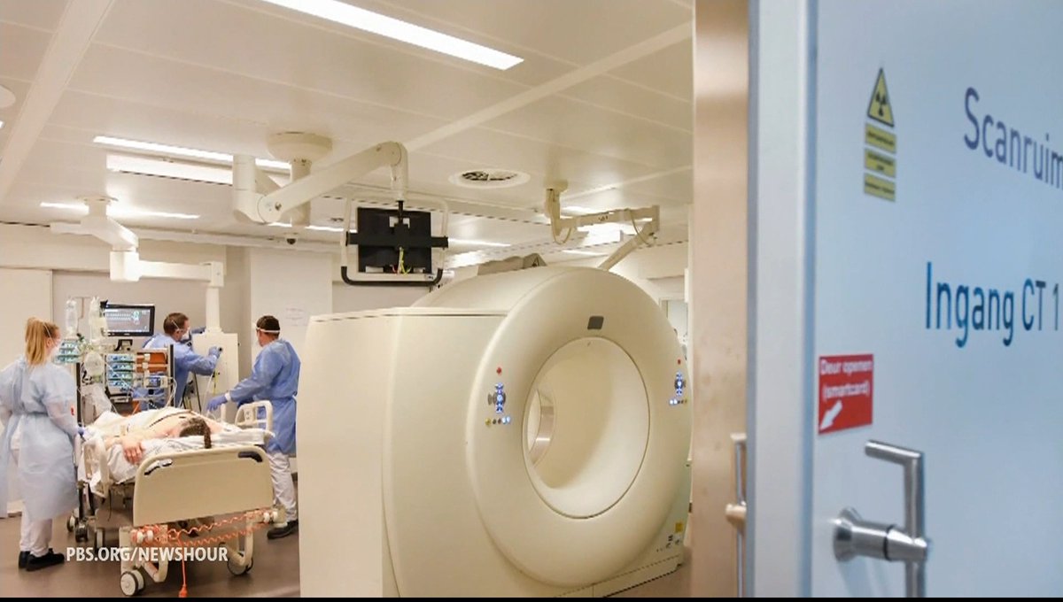
PHOTON COUNTING CT is NOT a better CT It is a NEW imaging modality Photon Counting CT (PCCT) represents a transformative leap in medical imaging, not only as a molecular imaging modality but also as a technology offering ultra-high resolution and functional imaging capabilities. It is fundamentally more than just an enhanced version of traditional CT—PCCT introduces new ways of seeing and understanding the human body, providing critical insights at the molecular, structural, and functional levels. This positions PCCT as a unique imaging modality that requires a fresh approach to technical implementation, operational workflows, and financial planning. Despite the larger upfront investment, PCCT’s ability to drastically reduce downstream healthcare costs makes it a highly valuable investment in the long run. 1. Technical Innovations • Molecular Imaging and Energy Discrimination: Unlike traditional CT, which simply measures the total absorbed energy, PCCT counts individual X-ray photons and differentiates their energy levels. This allows for precise molecular imaging, revealing the composition of tissues and materials at a biochemical level. By distinguishing between different tissue types and contrast agents, PCCT opens up new diagnostic possibilities, such as identifying molecular biomarkers in tumors or distinguishing between stable and unstable plaque in coronary arteries. This capability shifts the focus of imaging from purely anatomical to both anatomical and molecular, offering more comprehensive diagnostic information. • Ultra-High Spatial Resolution: PCCT features significantly smaller detector elements compared to conventional CT scanners, allowing for ultra-high resolution imaging. This means clinicians can visualize fine structures such as microcalcifications in arteries, small lesions in soft tissues, or the intricate architecture of bones. This level of detail was previously unattainable with traditional CT. When combined with molecular imaging, this ultra-high resolution allows for the precise localization and characterization of disease at very early stages, which is essential for early diagnosis and intervention. • Functional Imaging Capabilities: PCCT also excels as a functional imaging modality. By capturing energy-resolved information, PCCT can provide insights into tissue functionality and dynamic physiological processes. For instance, it can detect changes in blood flow, tissue perfusion, and oxygenation without the need for additional contrast agents or scans. This functionality allows for real-time assessment of physiological processes, making it particularly valuable in cardiology, oncology, and neurology for evaluating organ function and monitoring disease progression. • Reduced Noise and Artifact Reduction: Photon-counting technology dramatically reduces electronic noise and imaging artifacts, such as beam hardening, resulting in clearer and more accurate images. The ability to deliver ultra-high resolution images with minimal artifacts improves diagnostic accuracy, reducing the need for repeat scans and ensuring that even subtle abnormalities are detected. 2. Operational Considerations • New Workflow for Molecular, High-Resolution, and Functional Imaging: The integration of molecular, ultra-high resolution, and functional imaging into routine clinical workflows introduces complexity that requires adaptation. Radiologists and technicians need specialized training to interpret and analyze multi-energy datasets that include molecular and functional information. PCCT produces a vast amount of detailed data, requiring clinicians to adopt new imaging protocols and refine their diagnostic approaches to fully leverage its capabilities. • Post-Processing and Data Management: PCCT generates richer, more complex datasets, which necessitates advanced post-processing tools and data management systems. Existing PACS and imaging software may not be equipped to handle such large volumes of data or to process functional and molecular information effectively. This means healthcare institutions must invest in robust IT infrastructure, including upgraded software and storage solutions, as well as provide additional training for staff on new imaging analysis techniques. • Revised Clinical Protocols: The molecular, functional, and ultra-high resolution imaging capabilities of PCCT will likely prompt changes in clinical protocols. For instance, the need for contrast agents may be reduced, simplifying patient preparation and decreasing the risk of adverse reactions. Additionally, the ability to monitor physiological functions in real-time through functional imaging could lead to more dynamic diagnostic procedures, such as assessing the effectiveness of interventions or treatments in real-time. 3. Financial Impact • Higher Initial Investment: PCCT systems are more expensive than traditional CT scanners due to their advanced technology, which includes photon-counting detectors and the computational power required for high-resolution, molecular, and functional imaging. While this upfront cost is significant, it is crucial to view it in the broader context of the downstream benefits and cost reductions that PCCT offers. • Downstream Cost Reductions: Although the initial capital investment is higher, PCCT’s ability to combine molecular, functional, and ultra-high resolution imaging leads to substantial reductions in downstream healthcare costs. Its superior diagnostic accuracy minimizes the need for follow-up tests, repeat scans, or invasive diagnostic procedures, such as diagnostic coronary angiographies. For example, in cardiology, PCCT can precisely differentiate between types of coronary plaque, reducing the need for invasive procedures to assess risk. • Lower Overall Healthcare Expenditures: By enabling earlier, more accurate diagnoses, PCCT can reduce the overall cost of patient care. Early detection of disease, particularly through its molecular and functional imaging capabilities, allows for more targeted treatments, potentially preventing the need for more aggressive and expensive interventions down the line. For instance, early-stage tumor detection via molecular imaging could lead to less invasive treatments, reducing hospital stays and improving patient outcomes, ultimately driving down healthcare costs. • Increased ROI Through Enhanced Patient Outcomes: Over time, the combination of molecular, functional, and ultra-high resolution imaging enhances diagnostic precision, which translates into better patient outcomes. Improved diagnostic accuracy reduces the incidence of unnecessary procedures, minimizes treatment delays, and results in more personalized and effective care. This leads to increased patient satisfaction, better healthcare outcomes, and greater patient throughput—all factors that improve the institution’s return on investment (ROI). • Competitive Advantage and New Revenue Streams: By adopting PCCT, healthcare institutions position themselves at the forefront of advanced imaging technologies. The ability to offer molecular, functional, and ultra-high resolution imaging creates a competitive advantage, attracting more complex and high-value cases. This can boost the institution’s reputation for excellence in diagnostics, leading to increased referrals, new patient populations, and expanded revenue opportunities. Summary Photon Counting CT (PCCT) is not just an evolution of existing CT technology—it is a molecular, ultra-high resolution, and functional imaging modality that fundamentally transforms the diagnostic landscape. Its ability to capture detailed molecular data, visualize minute anatomical structures with ultra-high resolution, and provide real-time functional imaging opens new possibilities for earlier and more precise diagnoses. While the financial investment in PCCT is larger, the reduction in downstream healthcare costs through improved diagnostic accuracy, fewer unnecessary interventions, and earlier disease detection far outweighs the initial expense. For institutions committed to advancing patient care and improving long-term financial outcomes, PCCT is an essential investment in the future of medical imaging. The video attached shows a patient accessing the Hospital for ACS. PCCT can provide ALL the imaging information of the concurrent imaging modalities (CXR, CAG, Echo, CMR) that you see around it... that's a lot! #PhotonCountingCT #MolecularImaging #UltraHighResolution #FunctionalImaging #FutureOfImaging #AdvancedMedicalImaging #EarlyDiseaseDetection #InnovativeCT #CuttingEdgeHealthcare #PrecisionDiagnostics #HealthcareInnovation #MedicalTechnology #CostEffectiveImaging #NextGenCT #PatientCareRevolution

Dr. Filippo Cademartiri

11,849 Aufrufe • vor 1 Jahr

Announcing a new Coursera course: Retrieval Augmented Generation (RAG) You'll learn to build high performance, production-ready RAG systems in this hands-on, in-depth course created by and taught by , experienced AI and ML engineer, researcher, and educator. RAG is a critical component today of many LLM-based applications in customer support, internal company Q&A systems, even many of the leading chatbots that use web search to answer your questions. This course teaches you in-depth how to make RAG work well. LLMs can produce generic or outdated responses, especially when asked specialized questions not covered in its training data. RAG is the most widely used technique for addressing this. It brings in data from new data sources, such as internal documents or recent news, to give the LLM the relevant context to private, recent, or specialized information. This lets it generate more grounded and accurate responses. In this course, you’ll learn to design and implement every part of a RAG system, from retrievers to vector databases to generation to evals. You’ll learn about the fundamental principles behind RAG and how to optimize it at both the component and whole-system levels. As AI evolves, RAG is evolving too. New models can handle longer context windows, reason more effectively, and can be parts of complex agentic workflows. One exciting growth area is Agentic RAG, in which an AI agent at runtime (rather than it being hardcoded at development time) autonomously decides what data to retrieve, and when/how to go deeper. Even with this evolution, access to high-quality data at runtime is essential, which is why RAG is a key part of so many applications. You'll learn via hands-on experiences to: - Build a RAG system with retrieval and prompt augmentation - Compare retrieval methods like BM25, semantic search, and Reciprocal Rank Fusion - Chunk, index, and retrieve documents using a Weaviate vector database and a news dataset - Develop a chatbot, using open-source LLMs hosted by Together AI, for a fictional store that answers product and FAQ questions - Use evals to drive improving reliability, and incorporate multi-modal data RAG is an important foundational technique. Become good at it through this course! Please sign up here:

Andrew Ng

124,656 Aufrufe • vor 1 Jahr

This Zimbabwean member of parliament whose parliamentary submission has gone viral is making a valid point based on peer-reviewed research. She is essentially saying that men who have regular sex are also reducing their risk of prostate cancer. Medical studies suggest a link between frequent ejaculation and a reduced risk of prostate cancer. However, the exact number of times a man should ejaculate per month to maintain optimal prostate health has not been definitively established. A study published in the Journal of the American Medical Association (JAMA) found that men who reported ejaculating at least 21 times a month had a lower risk of developing prostate cancer compared to those who ejaculated less frequently. The study, which followed over 31,000 men for several years, suggested a correlation between frequent ejaculation and a lower risk of prostate cancer. A study conducted by Australian researchers and published in the journal British Journal of Urology (BJU) in 2003 found that men who ejaculated more than five times per week in their 20s had a reduced risk of prostate cancer compared to those who ejaculated less frequently. A 2008 study in the British Journal of Urology International examined over 30,000 men and found that those who reported higher ejaculation frequency showed a lower risk of prostate cancer. A 2016 study published in the journal European Urology investigated the association between ejaculation frequency and the risk of prostate cancer. The study found that men with a higher ejaculation frequency had a lower risk of developing prostate cancer, particularly in their 40s. Men over 40 should have regular medical check-ups and discuss any concerns about their prostate health with healthcare providers such as nurses and doctors. While this data is available for us to engage with, we cannot return to the old tradition she mentions in her presentation of passing on young women from the wife’s family to older men. The JAMA study's findings do not justify the revival of the outdated and exploitative traditional practice of passing young women from the wife's family to older men. Read more here; More👇🏿

Hopewell Chin’ono

127,956 Aufrufe • vor 1 Jahr