Loading video...

Video Failed to Load

Go Home

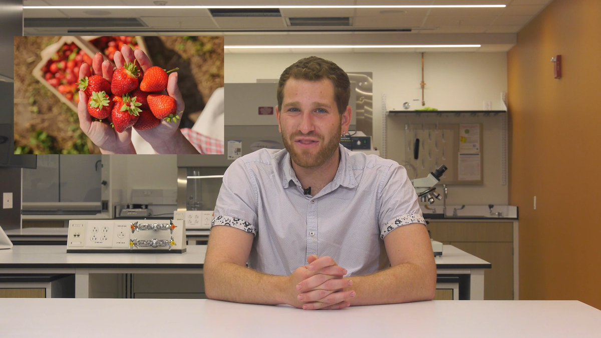
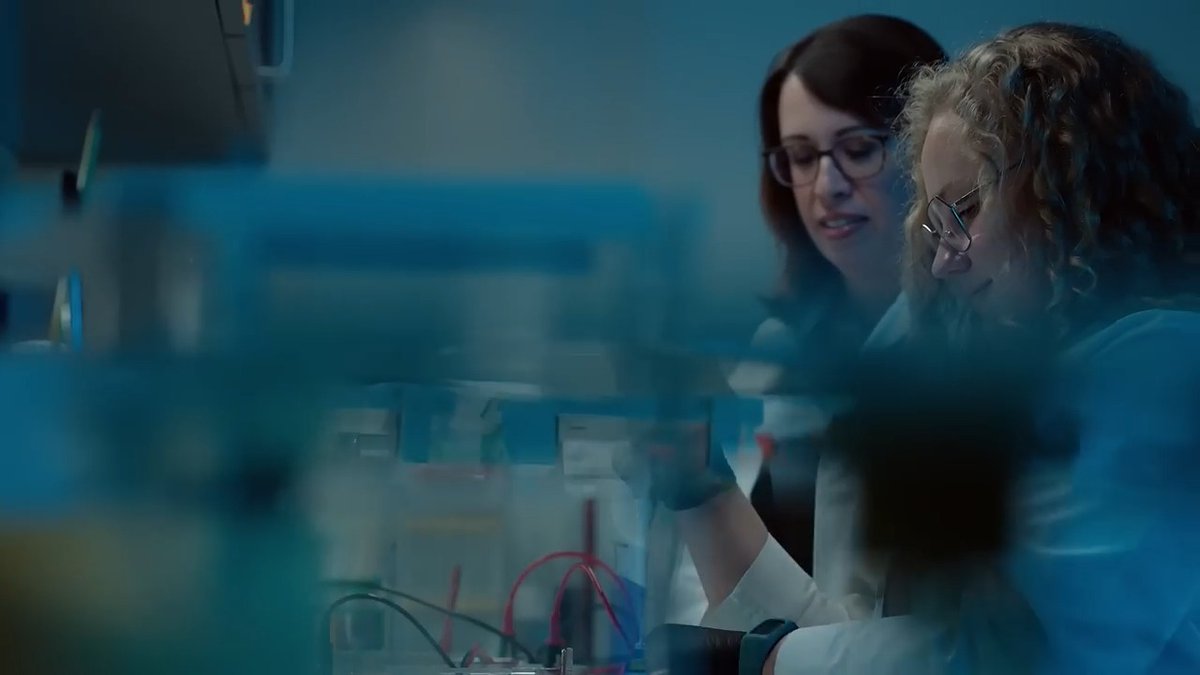
🧬 Take a look inside a 3D tissue bioprinting lab! NIH postdoctoral fellow Dr. Cristina Antich Acedo demonstrates how cutting-edge bioprinting can recreate human tissue structure. By combining different cell types and biomaterials, researchers can mimic real biological environments with precision. 🔬 This technology helps accelerate the path from...

14,495 views • 3 months ago •via X (Twitter)

0 Comments

No comments available

Comments from the original post will appear here

Related Videos

NEW episode! Drug development has never been more expensive, in terms of output per dollar spent. This trend, called Eroom’s law, is surprising, considering the incredible technological advances in drug discovery, from genome sequencing to engineering to microscopy. On a new episode of the Works in Progress podcast, Ben Southwood and I talk to Ruxandra Teslo 🧬 about why this has happened and what can be done about it. We discuss how: • AI isn’t a magic bullet for drug discovery. Predictive models lack the physical human data, like individual variation and rare side effects, that can only be generated by actually running real-world clinical trials. • As scientists invent more effective drugs, it becomes harder to discover new treatments that can surpass past successes. This is known as the "Better than the Beatles" problem. • Biotech companies are increasingly moving their "first-in-human" trials to Australia because its simpler regulations allow researchers to test drug safety faster and cheaper than in the US. • Clinical trials can be made more efficient with various reforms including: embracing platform trials, allowing researchers to select from independent ethics boards, expanding the funding and validation of surrogate endpoints, increasing transparency by releasing regulatory correspondence from failed companies, and much more. Timestamps: 00:00:00 Eroom’s law and the paradox of drug development 00:08:03 How clinical trials actually work 00:10:23 The power and controversy of surrogate endpoints 00:14:01 How historical patent laws influenced trial timelines 00:22:46 The Australia advantage and regulatory drag 00:29:08 Institutional review boards (IRBs) and bureaucratic drag 00:32:21 Open science and successful reforms 00:41:49 Our wishlist for clinical trial reforms, and which reforms we *don’t* like 00:53:48 Why AI isn’t a magic bullet for drug discovery

Saloni

109,256 views • 4 months ago

Dr Anthony Fauci authorized over $200 million US taxpayer dollars for transgender animal testing and animal torture “Can you describe what exactly the American people's taxpayer dollars were spent on regarding transgender animal testing?” “Yeah: - In a lot of these cases, they involve mice, rats, monkeys who are being surgically mutilated and subjected to hormone therapies to mimic female to male or male to female gender transitions - Gender affirming hormone therapies, and then looking at the biological, psychological, and physiological effects of the gender transitions - Looking at the effects of taking vaccines after you've transitioned these animals from male to female or female to male - Looking at the size of their genitals changing after you've put them on estrogen or testosterone therapies to transition them - There was a $1.1 million grant to give female lab rats testosterone to mimic transgender male humans and then overdose them with this party drug to see if female animals taking testosterone were more likely to overdose on the sex party drug than animals who are not taking testosterone “Are many of these taxpayer funded animal studies shared with the public or is there a significant oversight of this research?” “You essentially needed a degree in information technology to navigate the federal spending databases to find any of this stuff.” “In our analysis, Dr. Fauci funded about 95% of the change-drender animal experiments” “What we are doing to test human effects is poisoning lab animals - Forcing them to breathe wildfire smoke simulated in a laboratory by burning different types of foliage and pumping into animals cages - Making them obese to simulate what it would be like for obese people to be exposed to wildfire smoke - Shooting off handguns and rifles and forcing animals to breathe the emissions and gun control experiments - and the list goes on and on”

Wall Street Apes

237,297 views • 1 year ago

Excited to share our new work on building a multimodal atlas of human skin in health and inflammatory disease — a project I’m especially proud of, bringing together AI, high-throughput genomics, and clinical science to accelerate discovery. Over the past decade, single-cell genomics has transformed how we map cells in human tissues. But a major challenge remains: can we systematically decode how cells organize into functional niches in situ — including those invisible to standard histopathology? To address this, we integrated large-scale scRNA-seq, spatial transcriptomics, histopathology, and AI-driven modeling frameworks to build an in situ atlas of human skin across health and disease. Led by Lloyd Steele, an MD/PhD student working between Haniffa Lab and my lab at Wellcome Sanger Institute and Cambridge University . Another amazing collaboration with Muzz Haniffa, the mastermind behind the work as part of Human Cell Atlas. A key part of this study is that we didn’t build everything from scratch — we leveraged and combined AI methods that actually work! and showed how they can be used together to extract biological insight at scale. We used: • scArches to build and map into a reference scRNA-seq atlas of human skin: • NicheCompass to identify and characterize spatial niches: • MINT-Flow to extract microenvironment-induced cell states and gene programs: Together, these enabled an end-to-end workflow from atlas construction to spatial mapping, niche discovery, and cell state decoding. At scale, we integrated ~5 million cells and 100+ spatial sections, enabling a systematic view of tissue organization. Using this framework, we identified 26 niches in skin, including known histopathologic structures as well as hidden disease-associated niches not visible on H&E. Among the most striking findings were a resident memory T cell-rich sebaceous gland niche and a plasma cell-rich sweat gland niche, suggesting that appendageal structures act as active immunological microenvironments and may contribute to inflammatory memory and disease persistence. Importantly, this atlas is not just descriptive — it is usable. It can support mapping of new datasets, resolve finer cell types and niches, extract microenvironment-driven programs, and enable predictive analyses at scale. More broadly, this work shows what becomes possible when AI, spatial genomics, and atlas-scale data are integrated end-to-end: not just mapping tissues, but systematically decoding them. This was a massive collaboration, and I’m very grateful to the amazing scientists April Foster, Kenny Roberts, and Chloe Admane. Lloyd is an amazing scientist, and I’m especially excited for the community to see more of his work soon — stay tuned. The data and pre-trained models will be released soon. Preprint:

Mo Lotfollahi

11,819 views • 4 months ago

Excited to share TERRA, a tissue world model 🧬 Over ~1.5 years we ran a large data-generation + modelling effort to build a world model for human tissues, pretrained on 112M cells from spatial transcriptomics (mostly Xenium 5000-plex + public data). It's built on one of the largest human spatial transcriptomics corpora assembled to date, spanning 20 tissues across development, health and 26 disease conditions, ~two-thirds newly generated in-house. Why a "world model" for tissue? Images have universal representations (ViT/DINOv3), so do proteins (ESM, Alex Rives) and pathology (UNI, Faisal Mahmood). We've worked hard to build something similar for human tissue: one model that captures its multi-scale logic, genes → cells → their native microenvironments. Like the JEPA approach Yann LeCun has championed, TERRA learns by prediction in embedding space, but for human tissue. How it works: it tokenises each cell together with its nearest neighbours into one sequence while keeping every gene's identity, then masks part of a neighbourhood and predicts the representation of the hidden part, not raw noisy counts. From one backbone it reads out three scales, gene embeddings (what a gene is doing in a cell and its niche), cell embeddings (cell type and state) and neighbourhood embeddings (the niche), and because it keeps gene-level resolution it can knock a gene out in silico and predict the response. Applied entirely zero-shot, TERRA maps and perturbs human tissue across unseen organs, diseases and technologies, outperforming existing spatial approaches. Three take-homes: 1️⃣ One model, any tissue. A single pretrained backbone provides tissue representations zero-shot, handling genes, cells and niches across organs and platforms, off the shelf. 2️⃣ New biology, development to clinic. We built a new spatial atlas of the developing human pancreas and found an islet-associated capillary state that looks like a precursor of mature islet vasculature. In kidney, TERRA's in silico knockouts predicted the tissue-injury programme from cancer immunotherapy (checkpoint blockade), confirmed in treated kidneys, detected in blood, and linked to declining kidney function. 3️⃣ A grammar of tissue architecture. By coupling each cell's state to its niche, TERRA defines recurring cross-organ "archetypes" of macrophage neighbourhoods, including a tumour-boundary niche that tracks poor survival in kidney cancer. TERRA is already in use: it powered our recent skin atlas of hidden immune-memory niches ( with more studies coming soon. This was an amazing collaboration between clinicians, machine-learning scientists and cell biologists 🙏 Led by Sebastian Birk, Vali Sanian Amirhosein Vahidi, Samuel Ogden, Daniyal Jafree, Adib Miraki, Carlo Leonardi and Arpit Merchant, with Lassi Paavolainen, Menna Clatworthy, Omer Ali Bayraktar, Muzlifah Haniffa, Tom Mitchell and Mostafa Bakhti. Huge thanks too to everyone who shared data and helped along the way. What excites me most is seeing how the community builds on this. The model, code and tutorials are all public, so anyone can run TERRA on their own tissues, extend it, or build new models on top. Huge thanks to the whole team across Wellcome Sanger Institute and our many collaborators. 📄 Paper: 💻 Code: 🤗 Model: #SpatialTranscriptomics #SpatialGenomics #FoundationModels #AI4Science #MachineLearning #ComputationalBiology #SingleCell #WorldModels

Mo Lotfollahi

50,313 views • 18 days ago