

Niko McCarty.
@NikoMcCarty • 52,471 subscribers
Biotechnology for flourishing. Founding Editor @AsimovPress. Podcast, essays, occasional microgrants. Site: https://t.co/GabLJnLEnG Prev: Caltech, Imperial, NYU, MIT
Shorts


Videos
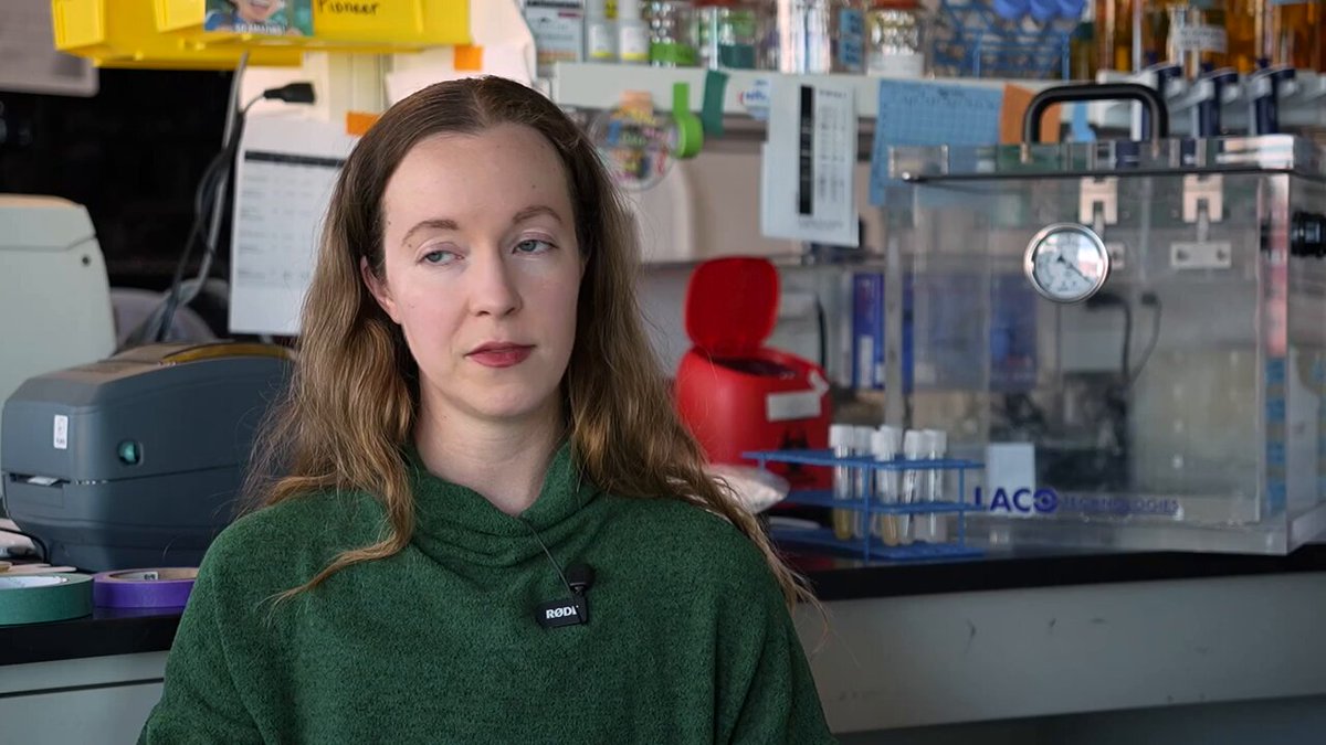
Breaking: PioneerLabs has engineered a microbe able to live on Mars, consuming solely nutrients found on the planet to create building materials. I interviewed Erika Alden DeBenedictis, the CEO, to learn how they did it and what might come next. Much of the interview was about bottlenecks to growing life on Mars; the planet has tons of water, but only in the form of ice. (If you melted all that ice, it would create an ocean 100 feet deep across the entire planet!) Similarly, we talked about the dirt. Martian regolith has salts, heavy metals, and perchlorates that are toxic to many organisms. Some microbes on Earth are able to break down these toxins, though, so Pioneer is engineering them to make them better at eating those toxins while adding nutrients back into the soil. This could possibly help support plant life in the future. Watch the full episode and subscribe to Asimov Press for more. 0:00 A first microbe engineered for Mars 5:03 Simulating Martian chemistry 8:27 The five "pioneer species" 17:20 Water, warming, and nitrogen 26:39 Radiation, growth rates, and poor measurements 35:49 Starship 41:05 Why create a second biosphere?
Niko McCarty.97,770 views • 26 days ago


Out today: Researchers have made the first cell capable of “feeding, growth, replication, division and selection...entirely using components scientists put there.” In other words, a cell that self-replicates and was made entirely from the ground-up, molecule by molecule. The cell is called SpudCell. And although it is definitely a cell (in that it has a membrane with molecules inside) it is definitely not alive, because it cannot grow indefinitely, survive without human help, make its own ribosomes, or recycle waste. It "dies" after a few divisions. But it's a starting point! And these same researchers have raised $6-8M+ in philanthropic funding to scale their efforts with a new nonprofit for synthetic cell research, called Biotic.
Niko McCarty.124,148 views • 3 months ago

New Episode: How to Design Flowers Nick Desnoyer uses genetic engineering, breeding, and pathogens to design and then create flowers that don't exist anywhere else on Earth. We talked about how he does it, in a room surrounded by many of his creations. At the start of the episode, Nick reveals his next project, called Paintunia, which is an engineered line of petunia that acts as a living canvas. He has engineered these petunias to include an inducible gene; when he lightly touches their buds with a molecule called dexamethasone, the molecules seep into cells, switch on the gene, and cause them to make a pigment. The result is a paintable flower, where molecules literally control which colors show up in particular places. Nick’s plan is ultimately to engineer these petunias to express three separate colors, for cyan, magenta, and yellow. Nick's ultimate dream is to build a botanical garden full of weird and wonderful flowers that exist nowhere else. We filmed this episode surrounded by flowers, and he patiently explained to me how each one gets its shape and pigmentation; how he combines breeding and genetic engineering to make new flowers; and how he searches scientific papers for mutations to figure out which plant crosses to make next. I think he convinced me that designing new kinds of flowers makes us look more closely at nature, and appreciate it more deeply. After all, all the genes and variations he uses in the laboratory are based on designs that have evolved elsewhere. Hope you enjoy!
Niko McCarty.11,547 views • 10 days ago


A few months ago, researchers made a synthetic cell that grows, eats, and divides. But it only grows for about five generations, and then it “breaks down” completely and stops moving. This is the reason it’s not yet considered “alive.” When I asked Kate Adamala, the lead author behind SpudCell, why she thinks this happens, she said something really simple and somewhat surprising: The cell cannot break down its own waste. In other words, it has all these proteins and RNA and DNA, but has no way to break them down and reuse the building blocks when they inevitably fail. So when a ribosome stops working, it just sits inside the cell and remains there for many generations. With each generation, more of the cell is filled up with broken pieces. Now, you could add a protease to these cells, but proteases are blunt-force instruments. They would destroy both working proteins *and* the broken ones, and so probably kill the cell. And when I asked Kate why we can't just use the enzymes and chaperones that natural cells use, she said there are too many of them. If you try to recreate all of biology's quality control processes, you end up with a hugely bloated metabolism inside what is supposed to be a minimal cell. The issue, then — the fundamental bottleneck — is that we do not have good tools to recognize WHAT is broken inside a cell and *then* destroy them while leaving healthy variants unscathed. My hunch here is that we need better molecular sensors. If we could distinguish between different forms of a protein, without destroying a cell entirely, then perhaps we could couple that recognition to a selective protease and recycle only the things that are broken. We also just don’t understand, at a high enough level, what happens when a ribosome breaks, and thus we can’t design good probes to sense those broken molecules. In trying to create a synthetic cell, then, we might also arrive at ideas for much better tools. Trying to create life is a really good way to figure out all the obstacles and stuff we don’t yet understand. But when Kate says she thinks they'll achieve perpetual cell division by 2030 (in the interview), I'm kind of skeptical. There's a lot of work to do here, and the tools we tend to use as biologists are often much broader than the precise, highly-evolved systems we find in nature.
Niko McCarty.11,388 views • 12 days ago


This video is one of the first times I thought biology was “cool.” It shows a neutrophil cell chasing a bacterium. Originally recorded in the 1950s by David Rogers at Vanderbilt University, the video gave me a deeper appreciation for life, even at the level of a single cell, because the neutrophil's movements seem so intentful, purposeful, aware. It wasn’t until recently, though, that I actually tried to demystify the neutrophil’s movements and understand how they happen. Here's what I learned: 1. The neutrophil's surface has thousands of protein receptors. Molecules secreted by the bacteria collide with these receptors. When that happens, the proteins change shape, slightly, and initiate a signaling cascade. 2. The neutrophil “knows” where to go because of a discrepancy in bound vs. unbound receptors. The side of the cell closest to the microbe will, probabilistically, have more "bound" receptors than the other side (because the molecules secreted by the microbe have a concentration gradient). This is how the neutrophil figures out which way to move. 3. Each bound receptor activates several G proteins located inside the cell membrane. Each G protein, in turn, switches on PI3K enzymes. In this way, the original signal is amplified; a single "activated" receptor might cause ~100 copies of PI3K to get switched on downstream. 4. The PI3K enzymes stick phosphates onto lipids in the cell membrane. The side of the neutrophil facing the bacterium now has more phosphates than the "back" side. Phosphate-binding proteins, such as GEF, accumulate and then recruit Rac, thus activating it. Rac, in turn, acts like a molecular switch, ultimately recruiting Arp2/3. (TL;DR: A bunch of proteins get activated, and the high phosphate concentration at the leading edge is the key signal for all this.) 5. At any given moment, the neutrophil has millions of actin molecules. These are the proteins used to build the cytoskeleton. Half of the actins are already “assembled” into filaments, but the other half are just floating around. Arp2/3 acts as a nucleator, grabbing onto actin and then starting a new cytoskeletal branch. More actin is assembled at the leading edge (where the Arp2/3 has accumulated), where they each push on the cell membrane with ~2 piconewtons of force. Hundreds of actin chains, pushing together, causes the cell to form protrusions. 6. The assembling actin chains push the cell at a speed of ~20 micrometers per minute (the length of about ten E. coli cells placed end-to-end.) As all of this is happening, another signaling cascade, nucleated at the back end of the cell, is dismantling actin filaments and recycling them. All this happens over a span of about 30 seconds. Much of this process is invisible; what we see, instead, is "just" a cell chasing its prey. But that's the wonderful thing about biology: A singular observation is usually more than enough fodder for a lifetime of work. The well is deep. There is always more to learn.
Niko McCarty.127,486 views • 9 months ago

For decades, biologists argued that birds migrate using magnetic fields, and that cryptochrome proteins in their eyes help them figure out where to fly. But the evidence for this has always been flimsy. In 1972, Wolfgang and Roswitha Wiltschko captured migratory European robins, put them in cages, covered the cages with a blanket, and wrapped electric coils around them. Once the birds lost their view of the sky, they still tried to move, or "hop," in the direction of their normal migration route. But when the Wiltschkos flipped the magnetic field around, the robins started hopping in random directions. They took this as evidence that, somehow, birds migrate using a magnetic sense. (Not bad!) In 2000, three theoretical biophysicists in Illinois, Ritz, Adem, and Schulten, proposed a mechanism for that magnetic sense. They argued that when blue light hits a receptor in the eye, it might create something called a "radical pair," locking two electrons into a spin state that a magnetic field could then nudge. They suggested that cryptochromes, light-sensitive proteins in the retina, might be responsible. (Speculative!) And then, in 2021, researchers apparently confirmed this! They took cryptochrome proteins from a type of migratory robin and put them in a test tube. They shone blue light at the proteins and found that this light induced them to form that transient "radical pair" state. And finally, they showed that magnetic fields could change this photochemistry where, in the presence of a magnetic field, the proteins formed fewer of these radical pairs, because the field forced a larger fraction of the excited molecules to ‘collapse’ back to their resting state. The experiment they used to measure this, though, has almost nothing to do with biology. You literally isolate the proteins, bombard them with a blue laser and, after each pulse, measure how much light the proteins absorb. For the robin cryptochrome protein, adding a magnetic field changed the absorbance *slightly.* But here’s the caveat: All of this was done in vitro, in a test tube, in a water-based buffer (rather than a cell). And, even worse, the experiment was done at 5 degrees Celsius (waaaay colder than the bird’s normal temperature) and at a low pH. And even after all this tinkering, they still *barely* saw a magnetic response! The news media didn't report many of these caveats. I randomly pulled one story from Science News and another from the University of Oxford, and neither even mentioned that the researchers ran the experiment at low temperature. The Oxford writeup actually hyped the result up more than the paper itself; that author wrote: “…the authors think the proteins involved could be significantly more sensitive in their native environment. In cells in the retina, the proteins are probably fixed and aligned, increasing their sensitivity to the direction of the magnetic field. Moreover, they are also likely to be associated with other proteins that could amplify the sensory signals." (Wow!) When Andrew York and Maria Ingaramo, two scientist working at Calico, heard about these results, they grew skeptical and decided to search for magnetically-responsive proteins — that actually work under physiological conditions — on their own. And that search, in turn, gave rise to Nonfiction Laboratories, a company that not only has created such proteins, but is now trying to commercialize them to make magnetically controlled cancer therapies that are only active near a tumor. The clip below is from my podcast.
Niko McCarty.21,714 views • 1 month ago

The Magnetobiology Episode: A company in San Francisco, called Nonfiction Laboratories, is building proteins (like antibodies and enzymes) that can be controlled using small magnets. In this episode, co-founder Maria Ingaramo and scientific advisor Andrew York explain how they engineered a protein, MagLOV, that responds strongly to magnetic fields, why most prior attempts have failed to replicate, and how the mechanism of magnetically-controlled proteins actually works. They also get into the “dream” use cases, like cancer drugs that activate only at the tumor, which might have a lower toxicity inside the body. This podcast is made possible by Astera Institute. I'm happy with how this episode came out. I think my interviewing skills are improving, and I'm getting better at building up context throughout the episode. Enjoy! Search for "The New Biology" on YouTube, Spotify, and Apple Podcasts. Timestamps: 00:00 - Opening 00:54 — Introduction 01:35 — The dream 05:38 — Why magnets vs. light or ultrasound 10:05 — The physics 17:48 — On the name "magnetogenetics" 21:25 — Birds and cryptochromes 27:09 — Why is the field filled with so much junk? 29:51 — Adam Cohen's molecule 33:24 — Markus Meister’s debunking 38:06 — The experiment 46:22 — Finding the LOV domain 54:11 — Singlets, triplets, and cysteine 56:54 — What the magnet is actually doing 1:05:13 — The conformational-change red herring 1:12:46 — The Quantum Biology Institute 1:19:31 — Founding Nonfiction Labs 1:24:38 — How to convince skeptical investors 1:29:39 — What a magnetogenetic medicine might look like 1:38:50 — First clinical indications 1:45:12 — The regulatory path 1:48:01 — What the field needs 1:54:30 — Appendix: Whiteboard lecture
Niko McCarty.45,502 views • 4 months ago

A 3D structural model of an entire bacterial cell. Biology is the coolest thing ever.
Niko McCarty.170,610 views • 2 years ago

Our second book is now available for pre-order. 🎉 Embracing the book's technology theme, we did something special: We encoded the entire book into DNA & packaged it into stainless steel capsules, where it will live for tens of thousands of years. 1,000 copies were made... 🧵
Niko McCarty.103,081 views • 1 year ago

The Bitter Lesson for Biology — Adam Green on Scaling Laws for Virtual Cells In this episode, the founder of Markov Biosciences, Adam Green, explains the "bitter lesson" for biology, the idea borrowed from Richard Sutton that large unbiased datasets and the right training objective tend to outcompete models with hard-coded rules and human priors. We talk a lot about virtual cells, sources of bias in data, and his evidence that virtual cells pre-trained on plain observational data show clean scaling laws, getting monotonically better at predicting unseen perturbations as the models grow, and beating a state-of-the-art model built specifically for that task. Our interview was about 3 hours long, but cut to 1.5 hours. My main goal was to understand Adam's perspective on virtual cells and his goal to "solve biology." Search for "The New Biology" on YouTube, Spotify, and Apple Podcasts. Timestamps: 00:00 — Cold open 01:58 — Clinical predictions from a virtual cell 05:38 — What is a "virtual cell"? 08:01 — The problems with single-cell RNA-seq 11:31 — The urns analogy 19:54 — Observational vs. perturbation 23:29 — The bitter lesson for biology 29:06 — Geometric Plackett-Luce 38:27 — Ablations and loss function 47:23 — Cells as specimens 59:26 — Antibody-Drug conjugates 1:11:16 — Will we ever understand the cell fully?
Niko McCarty.28,362 views • 4 months ago

I've started writing my book: "Biology is a Burrito & Other Essays." It is an interactive and highly visual look into the beauty, speed, and complexity of a living cell. I'm planning to print hardcover books while serializing the essays online. The first essay is now available at This was inspired by Stewart Brand's latest book, "Maintenance of Everything," which he developed in serialized form with Works in Progress. One cool thing about that book was that he improved each chapter with reader comments before printing the physical copies! I'll be doing the same with this book. If you send me feedback that improves the text, I'll credit you online and in the final print version. You can also sign up to get email updates when a new essay launches. Hope you enjoy!
Niko McCarty.34,308 views • 5 months ago

My first podcast is out today with Mark Budde 🦕🏆, CEO of plasmidsaurus. Plasmidsaurus took whole-plasmid sequencing from $600 to $15 and turned a "boring" service company idea into a hugely successful company that now serves >70,000 scientists. We talked about what it means to build a "boring" company, whether the Plasmidsaurus idea could apply to other technologies (like CRISPR screens), and why Plasmidsaurus isn't expanding into China. This podcast is made possible by Astera Institute. 00:00 – Enter Plasmidsaurus 01:21 – Reducing sequencing costs by 40x 04:17 – Scaling Oxford Nanopore technology 06:29 – Venture capital and building a "boring" company 13:03 – Plasmidsaurus vs. traditional CROs 22:25 – On selling customer data 30:10 – Building a moat 37:08 – 50-minute results 39:26 – Logistics and the UPS partnership 48:42 – The Chinese market 50:20 – Playbook for new biotech founders 52:31 – The future of biological reasoning and AI You can find the podcast, THE NEW BIOLOGY, on YouTube, Spotify, and everywhere else.
Niko McCarty.29,522 views • 5 months ago


New book chapter: "How to Weigh a Cell." This chapter explains how scientists have weighed cells throughout history, often using simple equipment and back-of-the-envelope calculations. It has lots of interactives, so you can "repeat" the experiments directly in your browser.
Niko McCarty.23,637 views • 4 months ago
The bacterial flagellum looks like a simple tail, or whip. But it’s actually a rotating motor, and perhaps the most sophisticated protein complex nature has ever evolved. In e. coli, these motors are capable of astonishing speeds; about 15,000 rpm. (The world record, according to one study, is for a Vibrio cell that was “clocked at 100,000 rpm by laser microscopy.) The flagellum propels the cell forward at speeds of 20-30 microns per second, or roughly 15 body lengths per second. If scaled up to the size of a cheetah, E. coli would *nearly* be the fastest land organism. The darting movements of a microbe were first observed in 1676 by Antony van Leeuwenhoek, a Dutch cloth merchant. Antony was delighted by the motion of his “animalcules,” writing: “I must say, for my part, that no more pleasant sight has ever yet come before my eye than these many thousands of living creatures, seen all alive in a little drop of water, moving among one another, each several creature having its own proper motion.” But Leeuwenhoek did not see flagella. He assumed, rather, that these animalcules must be “furnished with paws” instead. Christian Ehrenberg would not properly describe flagella until 1836. But amazingly, all the way up until the 1970s, nobody actually knew how the flagellum spun! In 1973, there were two competing models people argued over: the helical-wave (bending) model and the rotating (corkscrew) model. The first model suggested that the flagellum whipped back and forth, side-to-side, to propel the cell like paddle. The corkscrew model suggested that the whole flagellum instead spins around like a screw. In 1974, the corkscrew model finally won out. For two separate studies, scientists affixed flagella to glass slides using antibodies, and watched as the cells spun around and around like corkscrews. And finally, in just the last year, high-resolution structures of the flagellum have revealed a LOT more about its intricate assembly. The tail is made from ~20,000 self-assembling copies of a single protein, called flagellin. A “driveshaft,” or rod, spins the tail and is itself made of 26 protein subunits. Each “motor” in E. coli consists of 11 stators, each of which is made from 7 proteins.(Other types of cells have even more stators, and swim with much higher torques.) The flagellum spins when protons flow into the cell through tiny channels in these stators; akin to water running through a turbine. Each proton makes a small part of the stator change shape and push against the rotor, nudging it forward one step. With dozens of stators working at once, these nudges quickly spin the propeller. I'm writing an essay for Asimov Press about this now, and am really enjoying learning about the flagellum and its history. It's an extraordinarily complicated structure, though, and has been a challenge to understand!
Niko McCarty.51,977 views • 1 year ago

There is a new paper in Science proposing a mechanism for how homing pigeons navigate on cloudy days. (Hint: It's magnetic fields.) As with many magnetobiology papers, though, I'm skeptical of their proposed mechanism. These researchers, from Germany, found that pigeons have macrophages in their liver that accumulate lots of iron. They confirmed this with staining and other analyses. These macrophages also tend to cluster near nerve fibers in the liver (this is important for later). Then they did a really interesting experiment: - Train 34 pigeons to fly a particular 19-kilometer route. Ensure they all do this well. - Split the pigeons into two groups: treatment and control. - Inject the treatment group pigeons with liposomes loaded with clodronate. The macrophages eat these liposomes, and then clodronate kills the cells and scatters the iron. - Release the pigeons, from both groups, on an overcast day (it's thought that pigeons use magnetic fields to navigate when there is no sun). - All of the control pigeons reached their destination within 70 minutes, but the treated pigeons scattered in random directions. - (Important control experiment: The treated pigeons, released on a sunny day, flew like normal and reached their destination.) - This is taken as evidence that ??? macrophages --> iron --> navigation ??? via magnetic fields. But the mechanism is fuzzy. This experiment is super interesting, and it's clear that the treated pigeons really are unable to home to their destination using a magnetic field. But I'm not entirely convinced by the mechanism these authors propose. The main claim is that these iron-loaded macrophages "align" in a magnetic field, and that they shift according to the bird's orientation so that it can fix its direction. These macrophages (somehow) send signals to the nerve fibers in the liver, which then pass the messages to the brain, which allows the bird to navigate. The news coverage for this story suggests how this might happen: "One idea is that as the bird shifts its position relative to Earth’s magnetic field lines, the ferritin changes orientation and tugs on the web of fibers within a macrophage, possibly triggering the release of signaling molecules." (All you need to do is read the 2016 Meister paper, from the images below, to understand why such a mechanism is physically dubious.) The problem, though, is that the authors show (in their own study) that the iron in these pigeons' livers only act as a stable magnet at super low temperatures, below about 12 degrees Kelvin (or -260 degrees Celsius). At normal, physiological temperatures, the iron would be scrambled by the thermal motions of the tissue. Every measurement in the paper is taken at cryogenic temperatures, but a bird's body temperature is much higher, which means heat would likely destroy any magnetic alignment. The authors claim that MILLIONS of iron particles in the liver are all acting together to escape this effect, but they don't demonstrate the mechanism convincingly at all. If this claim is true, why not take homing pigeons (control vs. macrophage-depleted) and then rotate a magnetic field around them? You could record their neurons to see if there is some kind of signal coming from the liver.
Niko McCarty.22,441 views • 4 months ago

How to See Microbes from the Sky — Yonatan Chemla on Hyperspectral Biology Scientists have studied bacteria under microscopes for 400 years. But Yonatan Chemla, a postdoctoral fellow at MIT, has developed a technology that lets us see microbes from a drone 100 meters in the air. In this episode, we discuss the emerging field of "hyperspectral biology," which uses hyperspectral cameras (first built by NASA in the 1980s) to detect molecules with unique light-absorption signatures. With this technology, you could engineer a microbe to sense a landmine, for example, and release a pigment in response. A drone could then spot that pigment from hundreds of meters away, thus identifying the locations of the landmines. By spraying other types of engineered microbes over a field, they could similarly map heavy metals, report on soil health, or find gold deposits. But almost none of this can happen in the United States. The EPA regulates engineered microbes as chemicals under the Toxic Substances Control Act, a 1976 law that never mentions biology, and the agency seems to have approved just one product through it. As a result, many of biotechnology's most useful ideas — microbes that break down plastic, sense landmines, or remove pollution from water — never leave the laboratory. This episode is much more casual than other podcasts I've recorded. We talked for many hours, into the early morning, and so I also feel like my lines of questioning are not as good as it could've been. The video quality is also not great; sorry! Chapters: 00:00:00 Introduction 00:04:08 Applications for hyperspectral biology 00:07:33 How to build a biosensor 00:13:00 Molecular absorption data 00:22:12 How hyperspectral cameras work 00:38:15 Spatial resolution and satellite monitoring 00:51:04 Regulations around environmental release 01:08:44 Risk vs progress
Niko McCarty.15,832 views • 3 months ago