Video yükleniyor...

Video Yüklenemedi

Ana Sayfaya Dön

Inside every cell, there is a transport system that determines how materials move, signals propagate, and structure is maintained. This video captures that system in motion. The glowing streaks mark the growing ends of microtubules (protein polymers that constantly assemble and disassemble through a process called dynamic instability). Rather...

18,469 görüntüleme • 4 ay önce •via X (Twitter)

0 Yorum

Yorum bulunmuyor

Orijinal gönderinin yorumları burada görünecek
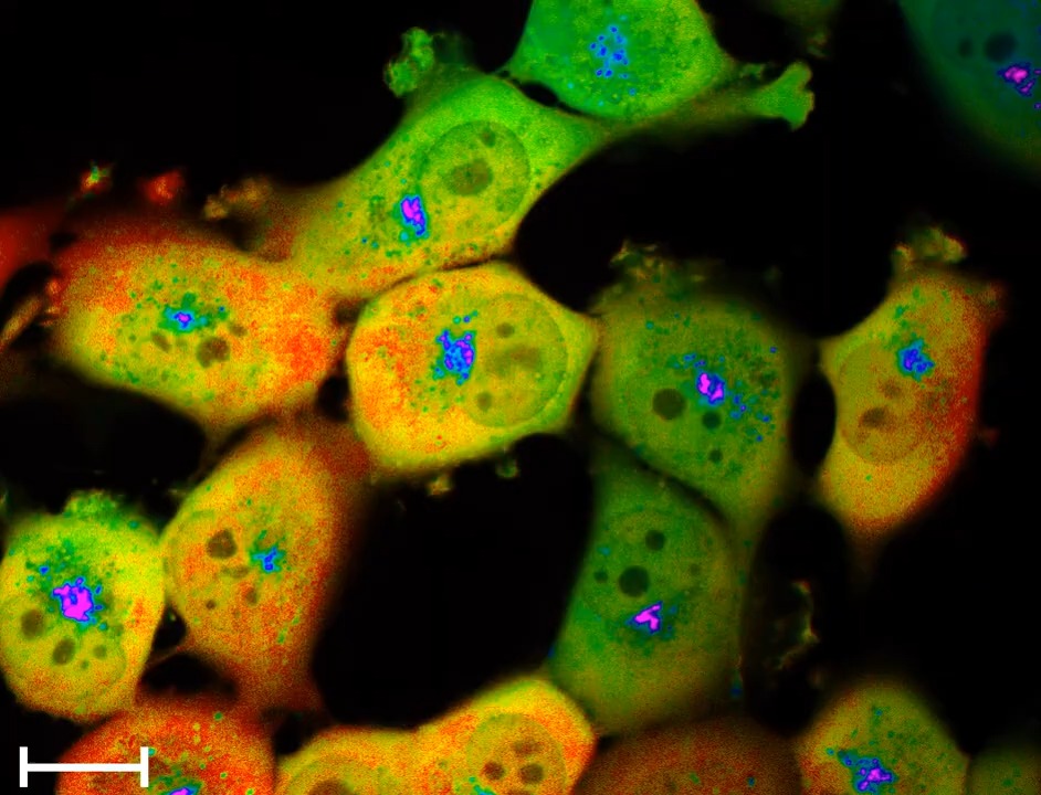
Benzer Videolar

🚨 Scientists discover wisdom teeth contain stem cells capable of repairing the heart, brain, and bones. Wisdom teeth contain dental pulp, a soft connective tissue threaded with blood vessels and nerves. Inside that pulp lives a dense population of mesenchymal stem cells, a class of undifferentiated cells that researchers classify as among the most therapeutically valuable biological material a human body produces. These are not ordinary cells maintaining routine tissue. They are blueprint cells, capable of receiving chemical signals from damaged environments and reshaping themselves into whatever the body needs most, neurons, cardiomyocytes, osteoblasts, even hepatic cells under the right conditions. The brain operates under a brutal rule: most of its neurons do not regenerate after damage. A stroke, a traumatic injury, a neurodegenerative disease removes cells the brain cannot replace through normal biological processes. Researchers have spent decades attempting to solve this through synthetic means, engineered cell therapies, growth factor injections, gene editing approaches that cost extraordinary resources and produce inconsistent results. What dental pulp stem cells demonstrated in laboratory conditions is that they can migrate toward neural damage sites, integrate with existing tissue architecture, and begin producing neurons and glial support cells. The mechanism involves neurotrophic factor secretion, essentially the cells releasing signaling proteins that stimulate the surrounding neural environment to repair itself from within. Cardiac muscle operates under a similarly unforgiving rule. After a heart attack, the dead muscle tissue becomes fibrotic scar material. The heart compensates by making surviving muscle work harder, a process that gradually leads to enlargement, weakening, and eventual failure. Dental pulp stem cells introduced into cardiac tissue in multiple studies produced measurable reductions in scar formation and demonstrated the ability to differentiate into functional cardiomyocytes, beating in synchrony with native heart cells. Some studies recorded improved ejection fraction in animal models, the core measurement of how effectively the heart pumps blood. Bone regeneration represents the most clinically advanced application already moving toward human trials. Dental pulp stem cells express high levels of osteogenic markers and respond rapidly to bone morphogenetic proteins, the chemical messengers that trigger skeletal repair. Their application in craniofacial reconstruction, spinal fusion, and long bone defect repair is being studied across multiple institutions simultaneously. What separates these cells from other stem cell sources is the combination of accessibility and biological youth. Bone marrow aspiration requires sedation and produces significant post procedure pain. Umbilical cord blood requires planning around birth. Wisdom teeth emerge between 17 and 25, during peak cellular vitality, and come out during a procedure most people already schedule. The extraction window is permanent. Once the teeth are gone and the pulp degrades, that specific population of young, highly potent cells is irretrievable from that individual. Cryogenic preservation protocols now exist that maintain dental pulp stem cell viability for over two decades. Several countries have commercial dental stem cell banks operating with the same institutional model as cord blood banking, long term frozen storage, indexed against future therapeutic need. The science supporting the value of preservation is no longer speculative. What lags behind is public awareness and clinical infrastructure in markets where this remains obscure. The wider pattern is worth recognizing. Medicine has repeatedly discovered that profound biological tools were present in tissues it previously categorized as vestigial, unnecessary, or inconvenient. The appendix was considered evolutionary junk for over a century before researchers identified its role in gut microbiome preservation. Wisdom teeth carried the same dismissal, a developmental relic from ancestors who needed extra molars for coarse diets, relevant only in their capacity to cause orthodontic problems. The pulp inside them was never junk. It was a repair system the body built during youth and stored in one of the most protected anatomical locations, surrounded by enamel, the hardest substance the human body produces. Evolution rarely wastes that kind of architecture.

The Curious Tales

24,267 görüntüleme • 3 ay önce

A single E. coli cell, placed on a dish, will become 70 billion cells in just 12 hours. That’s exponential growth. But a new preprint shows that it's possible to engineer E. coli to grow linearly instead, where only one daughter cell continues dividing and the other stops. First, some context. In nature, there is a bacterium called Mycobacterium smegmatis (initially discovered in 1884 in ulcers scraped from syphilis patients.) M. smegmatis is weird because it divides asymmetrically. These cells grow only from one end, and all their cell wall biosynthesis machinery is located on that one end. So when the cell divides, one daughter gets this machinery and the other gets nothing. The daughter that gets the machinery can keep dividing immediately, but the other daughter has to remake all that machinery from scratch, so its growth is delayed. E. coli doesn’t grow like this. When it divides, it pinches in the middle and splits everything evenly. Enzymes, metabolites, and proteins get partitioned more or less randomly between the two daughters. For the new preprint, though, researchers engineered E. coli to behave more like M. smegmatis. Here is how they did it: First, they deleted a gene called cyaA, which encodes an enzyme (adenylate cyclase) that makes a molecule called cAMP. cAMP is SUPER IMPORTANT! It is a nutrient sensor that instructs E. coli to switch on genes that help it digest non-glucose carbon sources when glucose is scarce. Without cAMP, E. coli cells growing on alternative carbon sources will starve; they won’t know how to eat the food. Next, they added back a “split” version of the cyaA gene into the cells. In other words, they split the gene in two so that each half of the enzyme is made separately. Cells can only make cAMP, and thus eat non-glucose carbon sources, if these two halves come together. To facilitate that “coming together,” the researchers also fused the split cyaA proteins to sticky proteins that clump together, and to a fluorescent protein (to make it easy to track these molecules in the cell.) So now some interesting things start to happen if you grow E. coli on a growth medium lacking glucose. As the cell grows, its cyaA “halves” start clumping together into a giant ball. Inside the aggregate, the two enzyme halves come together and make cAMP. And when the cell gets big enough and divides, the clump of cyaA RANDOMLY goes to either daughter cell #1 or #2. The daughter that gets the aggregate (called PA+ in this paper) can keep dividing. The daughter that doesn’t (PA–) cannot. It still grows a few times — about four divisions — because it inherits some leftover cAMP from its mother. But after that, the metabolite is diluted away, and the cell stops growing. PA+ cells went through about 23 divisions on average before their aggregate decayed. And the population of cells, as a whole, grew linearly. This paper is cool because there are many applications where exponential growth is too unpredictable and, perhaps, unsafe. If you want to engineer bacteria to deliver drugs, clean up waste, or live in the gut, you don’t want them to double uncontrollably. This paper shows you can make them expand in a controlled, linear way. Alas, mutations could break this whole engineered system. A mutation that restores cyaA, for example, would give cells a new way to make cAMP. Mutations that make the aggregates split between daughters would break the asymmetry, too. But still, I really enjoy proof-of-concept engineering papers like this.

Niko McCarty.

57,988 görüntüleme • 10 ay önce

There's a bacteriophage that turns bacteria into “liquid crystals.” Specifically, Pseudomonas aeruginosa bacteria make Pf phages, which are rod-shaped, negatively-charged, and measure about 2 micrometers in length (roughly the length of an E. coli cell). These phages leave the cells and enter their surroundings. There, they mix with polymers, also secreted by the cells, to form a crystalline matrix. Surprisingly, this is good for the cells. Although the phages kill some of them, it also makes their biofilms stickier and able to withstand certain antibiotics. These bacteria + phages are prevalent in cystic fibrosis patients; they've formed a sort of symbiotic relationship. The Pf phages are made from thousands of repeating copies of a coat protein, called CoaB, which wraps around a single-stranded, circular DNA genome. These genes are integrated directly on the bacterial chromosome. The bacteria “turn on” these phage genes when placed in a viscous environment with low oxygen levels. This is like a trigger to start forming a biofilm. And the cells make a lot of phages; about 100 billion per milliliter. These liquid crystals form because of a physics principle called “depletion attraction.” If you just mix a bunch of loose or flexible polymers together (such as long carbon chains) they will not form a liquid crystal. But if you mix stiff rods (the phages) with loose polymers at a high enough concentration, the polymers will force the phages close together to create a material that flows like a liquid despite being ordered like a crystal. See the video below. These liquid crystal biofilms are hard to get rid of. The negatively-charged phages block many antibiotics (like aminoglycosides, which are positively-charged) from entering cells. Liquid crystals also retain water, so these biofilms can survive on drier surfaces. I first heard about this from Malmesbury’s excellent newsletter, called “Telescopic Turnip.”

Niko McCarty.

50,024 görüntüleme • 5 ay önce

Some microbes carry a protein, called SNIPE, that "chops up" phage DNA as it's being injected into the cell. This is a new mechanism for phage defense! CRISPR–Cas and restriction enzymes also evolved to fight against phages, but they work by recognizing sequences. SNIPE works, instead, by sensing "touch." SNIPE is a protein with about 500 amino acids. After it's made by the ribosome, it latches onto ManYZ, two proteins which sit on the cell's inner membrane. (ManYZ is an importer; it brings mannose and other sugars into the cell.) Once attached to ManYZ, SNIPE sits and waits for an invading phage. Some phages, including lambda, actually infect cells by pushing their DNA through this ManYZ channel. Lambda uses its "tail" to reach inside the protein channel, basically, and inject its DNA. When this physical touch happens, though, SNIPE is waiting. As soon as the phage DNA starts entering the cell, and passes through ManYZ and SNIPE, it gets immediately destroyed. This means that SNIPE is the first phage defense system discovered, so far, that uses spatial positioning at the injection site to destroy invaders. But there are caveats, of course. If you untether SNIPE from ManYZ, such that it can freely diffuse through the cell, it will chew up the bacterium's genome. It is not a highly discerning nuclease! Also, SNIPE is not found in most bacteria. A prior pangenome study, which sequenced lots of different microbes, found that roughly a third of well-studied bacterial lineages had at least one member with a SNIPE-like protein. (For this paper, they just ported one of those homologs into an E. coli laboratory strain.) And finally, because SNIPE's mechanism is tightly tied to ManYZ, it cannot be used to defend against phages that enter the cell through different routes. T4 phages, for example, inject their DNA straight through the cell membrane and into the cytoplasm, without interacting with ManYZ. This is a nice basic science paper. Applications TBD. (Just remember that scientists figured out that bacteria had a phage defense system, called CRISPR-Cas, many years before it was repurposed into a gene-editing tool.) P.S. The video below shows how cells with the SNIPE gene (middle row) kill invading phages, and thus continue growing and dividing. Empty vector (top row) refers to bacteria carrying a plasmid with no SNIPE gene; this is a control group. And SNIPE E414A refers to cells which received a mutated SNIPE gene, where the glutamate at position 414 has been changed to an alanine, thus destroying the protein's nuclease activity. These cells also die when they get infected with a phage.

Niko McCarty.

20,321 görüntüleme • 3 ay önce

Today I received my 4th infusion of SGF (Stem Cell Growth Factors) at Edogawa Hospital in Tokyo, Japan. What is SGF? 🦷 SGF is derived from the dental pulp of children's naturally shed baby teeth. No stem cells are injected. Instead, the infusion contains the signaling molecules, growth factors, cytokines, and regenerative proteins that stem cells naturally produce. These biological messengers help coordinate communication between cells and are believed to support tissue repair, immune regulation, blood vessel health, and nerve regeneration. These growth factors are small enough to cross the blood brain barrier and reach the brain and nervous system directly. For someone like me with confirmed neuroinflammation, white matter atrophy, and small fiber neuropathy destroying my nerves from the inside out, the goal is remyelination. Rebuilding the insulation around my damaged nerve fibers.💯 Researchers have studied SHED (Stem Cells from Human Exfoliated Deciduous Teeth) for their regenerative potential in areas such as nerve repair, neuroinflammation, tissue healing, and age-related degeneration. Some people have nicknamed therapies like this the "Fountain of Youth" because the goal is not to replace damaged tissue, but to activate the body's own repair and regeneration pathways. Whether that nickname is deserved remains to be seen, but the science behind cellular signaling and regeneration is fascinating. Japan has become a global leader in regenerative medicine and allows access to therapies that are not currently available in the United States under its regenerative medicine framework. As Patient #27 in the McCairn–Edogawa Protocol, I am grateful to have the opportunity to experience this emerging science firsthand. 🧬🦷🇯🇵 #SGF #SHED #RegenerativeMedicine #StemCellScience #Neuroinflammation #SmallFiberNeuropathy #EdogawaHospital #Tokyo #MedicalInnovation

Heather C

28,478 görüntüleme • 16 gün önce